Anatomical directions and planes. Conditional division of the body of animals into parts and regions. Faculty of Veterinary Medicine and Biotechnology
The following planes are mentally drawn in the body of the animal (Fig. 10): longitudinal - sagittal and frontal and transverse - segmental.
Sagittal planes cut the animal's body from top to bottom, into right and left parts, and only one of them - the median sagittal plane - divides the animal's body into equal and symmetrical - right and left - halves; lateral sagittal planes divide the body of the animal into unequal and asymmetrical parts.
The frontal planes cut the body into upper, or dorsal, and lower, or abdominal, parts.
Segmental planes are drawn in the transverse direction and divide the body into transverse segments, or segments.
To further clarify the position of the organ and the direction of its parts (surfaces, edges, corners, etc.), the following topographic terms are used in anatomy: cranial - directed forward, towards the skull; caudal - directed towards the tail; lateral - directed to the side of the median sagittal plane; medial, directed back towards the median sagittal plane; dorsal - directed upwards in animals, towards the back; ventral - facing animals down, towards the abdomen.
Directions are indicated on the limbs: proximal - towards the body and distal - in the direction from the body.
On the thoracic and pelvic limbs, instead of the anterior surface facing forward, the term dorsal, or back, is used, for the opposite surface facing backwards - volar, or anti-back, on the thoracic limb, and plantar, or anti-back, on the pelvic limb.
BODY AREAS OF THE ANIMAL
In the body of the animal, the stem part and limbs are isolated (Fig. I). The stem part is divided into: head, neck, torso and tail. On the head, the brain and facial sections are distinguished. In the brain section, the following areas are considered: occipital, parietal, frontal, auricle, eyelids, temporal, parotid gland, laryngeal.
The facial section is divided into areas: nasal, nostril, infraorbital, upper lip, lower lip, chin, buccal, chewing muscle, submandibular.
The neck is subdivided into the nuchal region, the region of the brachiocephalic muscle, the tracheal region and the lower region of the neck.
The trunk includes the dorsal-thoracic, lumbo-abdominal and sacro-gluteal regions. The thoracic region is divided into the back and chest. The back is divided into the area of the withers and the dorsal area. On the chest, the right and left lateral thoracic regions are distinguished, as well as unpaired sternal and presternal regions.
The lumbar-abdominal region consists of the lumbar region, or lower back. On the abdomen, there are: the regions of the left and right hypochondrium, the region of the xiphoid cartilage, the right and left iliac regions, the right and left inguinal regions, the umbilical and pubic regions.
The sacro-gluteal region is divided into the sacral and gluteal regions.
Rice. 11. Areas of the body of the cow:
Brain section of the head. Regions: 1 - occipital; 2 - parietal; 3 - frontal; 4 - auricle; 5 - century; 6 - temporal; 7 - parotid gland; 8 - guttural.
The facial region of the head. Areas: a - nasal; 10 - nostrils; 11 - infraorbital; 12 - upper lip; is - lower lip; 14 - chin; 15 - buccal; 16 - chewing muscle; 17 - submandibular.
Neck. Areas: 18 - vynaya; 19 - brachiocephalic muscle; 20 - tracheal; 21 - lower region of the neck.
Dorsal-thoracic region. Areas: 22 - withers; 23 - dorsal; 24 - lateral chest; 25 - sternum; 26 - presternal.
Lumbar-abdominal. Areas: 27 - lumbar (lumbar); 28 - stomach.
Sacro-buttock department. Areas: 29 - sacral; 30 - gluteal. Thoracic limb. Regions: 31 - shoulder girdle, or scapula; 32 - shoulder; 33 - forearm; 34 - wrist; 35 - metacarpus; 36 - the first phalanx; 37 and 38 - the second and third phalanxes. Joints: 39 - shoulder; 40 - elbow; 41 - carpal; 42 - putovy (first phalanx); 43 - coronal (second phalanx); 44 - hoofed (third phalanx). Pelvic limb. Areas: 45 - pelvic girdle; 46 - groats; 47 - hips; 48 - knee cup; 49 - lower leg; 50 - tarsus; 51 - metatarsus; 52 - the first phalanx (outside the hooves); 53 - second phalanx; 54 - third phalanx. Joints: 55 - hip; 56 - knee; 57 - tarsal (hock); 58 - putovy (first phalanx); 59 - coronal (second phalanx); 60 - hoofed (third phalanx).
As part of the thoracic limb, the region of the shoulder girdle, or scapula, associated with the body, and the free thoracic limb are considered. The free thoracic limb is subdivided into the areas of the shoulder, forearm, wrist, metacarpus, first phalanx of the fingers, second phalanx of the fingers and third phalanx
In order to be able to navigate the body of the animal, indicate the topography of its individual organs and make it easier to study it, the body of the animal was divided into regions, departments, which received a specific name.
Along with the complication of the structure of the body of vertebrates, the conditional division of it into regions becomes more complicated.
In fish, the head, trunk (the area between the head and tail) and tail (the area located behind the anus) stand out on the stem part of the body.
In terrestrial vertebrates, in connection with the development of their limbs, two parts are already distinguished on the body - the neck and the body (therefore, the body means the part without the neck).
In this regard, the head, neck, trunk and tail stand out on the stem part of the body; on the limbs - belts and free limbs (Fig. 7).
HEAD - caput. It is subdivided into the skull - cranium and the face - fades.
For quick and clear orientation in determining the places of damage on the head or when taking measurements in breeding work, regions are distinguished on the skull - regiones (rg.): On the border between the neck and head, the occipital region - rg. occipitalis; in front of her on top of the parietal region - rg. parietalis; in front of the parietal region, the frontal region is rg. frontalis; on the sides of it the area of the auricle - rg. auricularis; between the eye and ear on the sides of the parietal region, the temporal region - rg. temporalis.
On the face, they distinguish - "the region of the nose - rg. nasalis, on which the back of the nose - dorsum nasi, the tip of the nose - apex nasi and the lateral region - rg. lateralis nasi stand out; on the sides and below the latter is the infraorbital region - rg. infraorbitalis, passing into buccal region - rg. buccalis, on which the maxillary, dental and mandibular areas are distinguished; behind the buccal - zygomatic region - rg. zygomatica; behind the buccal region, where the large flat chewing muscle is located, lies the chewing region - rg. masseterica.
Below the face, between the lower jaws, are the intermaxillary region - rg. intermandibularis and the area of the hyoid bone - rg. subhyoidea. On the anterior part of the face, its apical or apical part, the region of the nostrils is distinguished - rg naris, the region of the upper lip - rg. labialis superior. In the region of the nostrils and upper lip, there may be a nasal or nasolabial mirror. The pigs have a snout here. There is also an area of the lower lip - rg. labialis inferior and chin area - rg. mental is.
Around the eye - the orbital region - rg. orbitalis, on which the region of the lower eyelid is distinguished - rg. palpebral superios
Rice. 7. Areas of the cow's body
NECK - collum (cervix). It borders on the occipital region, on the sides of which lie: the region of the parotid gland - rg. paratidea, located below the auricle, passing from above into the behind-the-ear region - rg. retroauricularis, and from below - into the pharyngeal - rg. pharyngea; laryngeal region - rg. laryngea lies below behind the pharyngeal region. Along the lower side of the neck from the laryngeal region back to the body stretches the tracheal region - rg. trachealis. Along the neck from the sides of the tracheal region is the brachiocephalic muscle, the region of which is called the region of the brachiocephalic muscle - rg. brachiocephalica. Along the lower edge of this area stretches the jugular groove - sulcus jugularis, in which lies the external jugular vein, from which blood is usually taken from large animals. Below this gutter, the sternocephalic region is rg. sternocephalica; closer to the scapula, in the upper part it is called the prescapular region - rg. prescapularis. The back ventral part of the neck - dewlap - palear.
Above the region of the brachiocephalic muscle is the lateral cervical region, located in the upper part of the neck, - rg. colli lateralis, it still distinguishes the outer edge - margo nuchalis or the dorsal edge of the neck - margo colli dorsalis.
BODY - truncus. It distinguishes the dorsal-thoracic, lumbar-abdominal and sacro-gluteal regions.
The dorsal-thoracic region is a continuation of the bulge and upper regions of the neck, which consists of two parts: in front of the withers - rg. interscapularis and behind the dorsal region - rg. dorsalis.
On the sides and below from the back there is an extensive lateral chest region, from below passing in front of the presternal region - rg. presternalis, bordering on the tracheal, and behind - in the sternal - rg. sternalis.
The lateral thoracic region is also divided into two parts: the anterior part, where the shoulder girdle (scapula) lies on the chest, and the shoulder, which in many animals goes to the level of the sternal region. Caudal part of the thoracic region - costal - rg. cos-talis - reaches the edge of the chest, called the costal arch.
Lumbar-abdominal. The upper part of this department is the lumbar region - rg. The iumbalis (lower back) is an extension of the back. Below the waist - a vast abdominal region, or simply the belly (belly) - abdomen.
By two transverse (segmental) planes, drawn at the level of the most convex part of the costal arch and at the level of the maklok, the abdominal region is divided into three sections: the anterior region, in front and below, running along the edges of the costal arches (right and left) and posteriorly bounded by a transverse plane drawn along the edge of the convex part of the costal arch. This area is called the area of the xiphoid cartilage - rg. xiphoidea. The middle lateral region is located between the two transverse planes described above. Here are the right and left iliac regions - rg. iliacea. In this area, a hungry fossa (periolumbar fossa) fossa paralumbalis is distinguished, located under the lower edge of the lower back in front of the maklok, and the umbilical region - rg. umbilicalis - a site located in the middle region behind the region of the xiphoid cartilage (in this region the umbilical cord is located in newborns).
On the sides and behind the iliac region lie the right and left inguinal regions - rg. inguinalis, from below, as a continuation of the umbilical region, there is a pubic region - rg. publica.
Sacro-buttock department. In the middle part of this department, above and behind the lumbar region lies the sacral region - rg. sacralis, which passes into the root of the tail - radix caudae. On the sides of it is the gluteal region - rg. glutea, its lower border goes along the line passing from the maklok through the hip joint to the ischial tuberosity.
Gluteal region (buttocks) - rg. glutea (nates) is located in place of the pelvic girdle. Together with the sacral section, the paired gluteal region forms a croup in ungulate animals. The back side of the croup under the tail is called the anal region - rg. analis, here is the anus - the anus. Under the anal region from the anus to the labia in females and the scrotum in males lies the area perineum, or perineum, - rg perineals (perineum).
From the lower border of the gluteal region to the knee joint on the pelvic limb are the thigh - femur and the area of the patella - rg. patellaris, the knee fold rises up from it to the stomach. From the knee to the tarsal joint lies the lower leg - crus, from which the limb ends with a link called the foot - pes, or hind leg.
On the thoracic limb, the region of the shoulder girdle is distinguished - rg. scapularis (to the level of the shoulder joint) and the shoulder area - rg. brachials. These two areas are adjacent to the thoracic region. On the area of the shoulder girdle, another area of \u200b\u200bthe scapular cartilage is isolated - rg. suprascapularis, supraspinous - rg. supraspinata and infraspinal region - rg. infraspinata, located along the scapula in front of and behind the spine of the scapula.
From the shoulder joint to the elbow, there is a shoulder - brachium, behind which the edge of the triceps muscle, or the triceps edge, margo tricepitalis, is clearly visible. Between the elbow and wrist joints lies the forearm - antebrachium, below it is the hand - manus, or front paw.
Terms indicating the location and direction of parts of an animal's body. To clarify the location on the body of an organ or its part, the entire body is conditionally dissected by three mutually perpendicular planes drawn along the body, across and horizontally (Fig. 8).

Rice. 8. Planes and directions in the body
The vertical plane that cuts the body longitudinally from head to tail is called the sagittal plane - planum sagittate. If the plane passes along the body, dividing it into right and left symmetrical halves, then this is the middle sagittal plane - planum medianum. All other sagittal planes drawn parallel to the median sagittal plane are called lateral sagittal planes - plana of the sagittal plane, directed towards the median plane is called medial; the opposite (outer) area is called lateral, it is directed to the side. So, the outer surface of the rib will be lateral, and the one that is visible from the inner surface of the chest, i.e., towards the median sagittal plane, will be medial. The outer lateral surface of the limb is lateral, while the inner one, directed towards the median plane, is medial.
It is also possible to dissect the body with longitudinal planes, but in animals they are located horizontally on the earth's surface. They will run perpendicular to the sagittal. Such planes are called dorsal (frontal). These planes can be used to cut off the dorsal surface of the tetrapod body from the ventral surface. And everything that is directed to the back received the term "dorsal" (dorsal). (In animals it is upper, in humans it is posterior.) Everything that is directed to the abdominal surface has received the term "ventral" (abdominal). (In animals it is lower, in humans it is anterior.) These terms apply to all parts of the body, except for the hand and foot.
The third planes along which you can mentally dissect the body are transverse (segmental). They run vertically, across the body, perpendicular to the longitudinal planes, cutting it into separate sections - segments, or metameres. In relation to each other, these segments can be located towards the head (skull) - cranially (from Latin cranium - skull). (In animals it is forward, in humans it is up.) Or they are located towards the tail - caudally (from Latin cauda - tail). (In quadrupeds it is back, in humans it is down.)
On the head, directions are indicated towards the nose - rostral (from lat. Rostrum - proboscis).
These terms can be combined. For example, if it is necessary to say that the organ is located towards the tail and towards the back, then they use a complex term - caudodorsally. Both medical and veterinarian will understand you. If we are talking about the ventrolateral location of the organ, this means that it is located on the ventral side and outside, on the side (in the animal on the side - from below, and in humans on the side - in front).
In the region of the autopodia of the extremities (on the hand and foot), the back of the hand or the back of the foot are distinguished - dorsum manus and dorsum pedis, which serve as a continuation of the cranial surfaces of the forearm and lower leg. Opposite the dorsal on the hand are palmar (from lat. palma manus - palm), on the foot - plantar (from lat. planta pedis - sole of the foot) surfaces. They are called anti-back. In the region of the stylo- and zeugopodium, the anterior surface is called cranial, the opposite is called caudal. The terms "lateral" and "medial" are retained on the limbs.
All areas on the free limb in relation to their longitudinal axis can be closer to the body - proximally or further from it - distally. Thus, the hoof is located more distally than the elbow joint, which is located proximal to the hoof.
ANATOMY OF PETSPLANES OF THE BODY AND TERMS FOR DESIGNATION OF THE LOCATION OF THE BODY
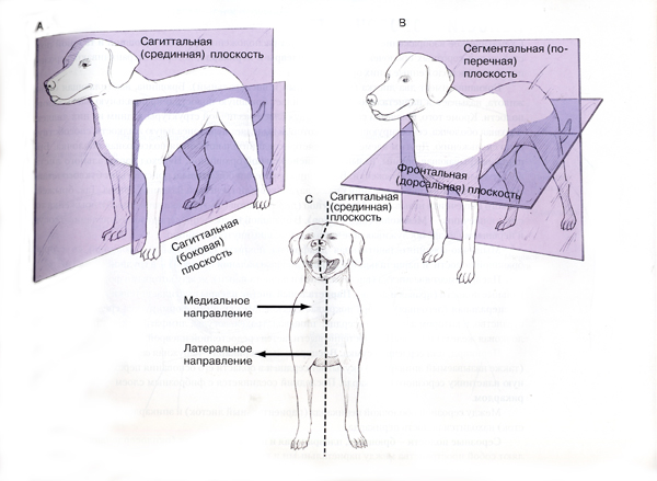
To determine the location of organs and parts, the body of the animal is dissected by three imaginary mutually perpendicular planes - sagittal, segmental and frontal (Fig. 1).median sagittal(median) plane is carried vertically along the middle of the animal's body from the mouth to the tip of the tail and cuts it into two symmetrical halves. The direction in the animal's body towards the median plane is called medial and from her lateral(lateralis - lateral).
^ Fig.1. Planes and directions in the body of an animal
Planes:
I– segmental;
II - sagittal;
III - frontal.
Directions:
1 - cranial;
2 - caudal;
3 - dorsal;
4 – ventral;
5 – medial;
6 – lateral;
7 - rostral (oral);
8 – aboral;
9 – proximal;
10 – distal;
11 – dorsal
(back, back);
12 – palmar;
13 - plantar.
segmental the plane is drawn vertically across the body of the animal. The direction from it towards the head is called cranial(cranium - skull), towards the tail - caudal(cauda - tail). On the head, where everything is cranial, they distinguish the direction towards the nose - nasal or proboscis - rostral and its opposite caudal.
Frontal the plane (frons - forehead) is drawn horizontally along the body of the animal (with a horizontally elongated head), i.e. parallel to the forehead. The direction in this plane towards the back is called dorsal(dorsum - back), to the stomach - ventral(venter - belly).
There are terms to determine the position of limb sections proximal(proximus - nearest) - a closer position to the axial part of the body and distal(distalus - remote) - a more distant position from the axial part of the body. To designate the anterior surface of the limbs, the terms cranial or dorsal(for the paw), and for the back surface - caudal, as well as palmar or volar(palma, vola - palm) - for the brush and plantar(planta - foot) - for the foot.
^
DEPARTMENTS AND AREAS OF THE ANIMAL BODY AND THEIR BONE BASIS
T 
The body of animals is divided into the axial part and limbs. Starting with amphibians, in animals the axial part of the body is divided into the head, neck, trunk and tail. The neck, trunk and tail are body stem. Each of the parts of the body is divided into sections and regions (Fig. 2). In most cases, they are based on the bones of the skeleton, which have the same names as the regions.
Rice. 2 ^ Areas of the body of cattle
1 - frontal; 2 - occipital; 3 - parietal; 4 - temporal; 5 - parotid; 6 - auricle; 7 - nasal; 8 - areas of the upper and lower lips; 9 - chin; 10 - buccal; 11 - intermaxillary; 12 - infraorbital; 13 - zygomatic; 14 - eye area; 15 - a large chewing muscle; 16 - upper cervical; 17 – lateral cervical; 18 - lower cervical; 19 - withers; 20 - back; 21 - costal; 22 - presternal; 23 - sternal: 24 - lumbar: 25 - hypochondria; 26 - xiphoid cartilage; 27 - lumbar (hungry) fossa; 28 - side area; 29 - inguinal; 30 - umbilical; 31 - pubic; 32 - maklok; 33 - sacral; 34 - gluteal; 35 - root of the tail; 36 - ischial region; 37 - scapula; 38 - shoulder; 39 - forearm; 40 - brush; 41 - wrist; 42 - metacarpus; 43 - fingers; 44 - hip; 45 - shin; 46 - foot; 47 - tarsus; 48 - metatarsus.
Head(Latin caput, Greek cephale) is divided into the skull (brain) and face (facial). The skull (cranium) is represented by regions: occipital (nape), parietal (crown), frontal (forehead) with the horn region in cattle, temporal (temple) and parotid (ear) with the auricle region. On the face (facies) there are areas: orbital (eyes) with areas of the upper and lower eyelids, infraorbital, zygomatic with the area of the large chewing muscle (in a horse - ganache), intermaxillary, chin, nasal (nose) with the area of nostrils, oral (mouth) , which includes the areas of the upper and lower lips and cheeks. Above the upper lip (in the region of the nostrils) there is a nasal speculum, in large ruminants it extends to the region of the upper lip and becomes nasolabial.
Neck
The neck (cervix, collum) extends from the occipital region to the scapula and is divided into regions: the upper cervical, lying above the bodies of the cervical vertebrae; lateral cervical (area of the brachiocephalic muscle), running along the vertebral bodies; the lower cervical, along which the jugular groove stretches, as well as the laryngeal and tracheal (on its ventral side). In ungulates, the neck is relatively long due to the need to feed on pasture. Fast gait horses have the longest neck. The shortest is in the pig.torso
The trunk (truncus) consists of the thoracic, abdominal and pelvic regions.^ Thoracic includes the areas of the withers, back, lateral costal, presternal and sternal. It is durable and mobile. In the caudal direction, strength decreases, and mobility increases due to the peculiarities of their connection. The bones of the withers and back are the thoracic vertebrae. In the region of the withers, they have the highest spinous processes. The higher and longer the withers, the greater the area of attachment of the muscles of the spine and the girdle of the chest limb, the more sweeping and more elastic the movements. There is an inverse relationship between the length of the withers and the back. The longest withers and the shortest back are in the horse, and vice versa in the pig.
^ Abdominal includes the lower back (lumbus), abdomen (abdomen), or belly (venter), therefore it is also called the lumbo-abdominal region. The loin is the continuation of the back to the sacral region. Its basis is the lumbar vertebrae. The abdomen has soft walls and is divided into a number of areas: the right and left hypochondrium, xiphoid cartilage; a paired lateral (iliac) with a hungry fossa, adjoining from below to the lower back, in front - to the last rib, and behind - passes into the inguinal region; umbilical, lying below the abdomen behind the region of the xiphoid cartilage and in front of the pubic region. On the ventral surface of the areas of the xiphoid cartilage, umbilical and pubic in females, the mammary glands are located. The horse has the shortest loin and a less extensive abdominal region. Pigs and cattle have a longer loin. The most voluminous abdominal region in ruminants.
^ Pelvic region(pelvis) is divided into areas: sacral, gluteal, including maklok, ischial and perineal with adjacent scrotal area. In the tail (cauda) distinguish the root, body and tip. The sacral, two gluteal, and root areas of the horse's tail form the croup.
limbs(membra) are divided into thoracic (anterior) and pelvic (rear). They consist of belts, which are connected to the stem part of the body, and free limbs. Free limbs are divided into the main supporting column and paw. The thoracic limb consists of the shoulder girdle, shoulder, forearm and hand.
Areas shoulder girdle and shoulder adjacent to the lateral thoracic region. The bone base of the shoulder girdle in ungulates is the scapula, which is why it is often called the scapula region. Shoulder(brachium) is located below the shoulder girdle, has the shape of a triangle. The bone base is the humerus. Forearm(antebrachium) is located outside the skin trunk pouch. Its bone base is the radius and ulna. Brush(manus) consists of the wrist (carpus), metacarpus (metacarpus) and fingers (digiti). In animals of different species, there are from 1 to 5. Each finger (except the first) consists of three phalanges: proximal, middle and distal (which in ungulates are called put, respectively, in horses - grandmother), coronal and hoofed (in horses - ungulates) .
The pelvic limb consists of the pelvic girdle, thigh, lower leg and foot.
Region pelvic girdle(pelvis) is part of the axial part of the body as the gluteal region. The bone base is the pelvic or innominate bones. Region hips(femur) is located under the pelvis. The bone base is the femur. Region shins(crus) is located outside the skin trunk pouch. The bone base is the tibia and tibia. Foot(pes) consists of a tarsus (tarsus), a metatarsus (metatarsus) and fingers (digiti). Their number, structure and names in ungulates are the same as on the hand.
^
SOMATIC SYSTEMS
The skin, skeletal muscles and skeleton, forming the body itself - the soma of the animal, are combined into a group of somatic systems of the body.
The apparatus of movement is formed by two systems: bone and muscle. The bones, combined into a skeleton, are a passive part of the apparatus of movement, being levers that are acted upon by the muscles attached to them. Muscles act only on bones that are movably connected with ligaments. The muscular system is the active part of the apparatus of movement. It provides the movement of the body, its movement in space, search, capture and chewing of food, attack and defense, breathing, eye and ear movements, etc. It accounts for 40 to 60% of the body mass. It determines the shape of the animal's body (exterior), proportions, determining the typical features of the constitution, which is of great practical importance in zootechnics, because. endurance, adaptability, fattening ability, precocity, sexual activity, vitality, and other qualities of animals are associated with the features of the exterior, the type of constitution.
^
SKELETON, CONNECTION OF BONES OF THE SKELETON (OSTEOLOGY)
General characteristics and significance of the skeleton.
The skeleton (Greek skeleton - withered, mummy) is formed by bones and cartilage, interconnected by connective, cartilaginous or bone tissues. The skeleton of mammals is called internal, because. it is located under the skin and is covered by a layer of muscle. It is the solid foundation of the body and serves as a case for the brain, spinal and bone marrow, for the heart, lungs and other organs. The elasticity and spring properties of the skeleton provide smooth movements, protect soft organs from shocks and tremors. The skeleton is involved in mineral metabolism. It contains large reserves of calcium, phosphorus and other substances. The skeleton is the most accurate indicator of the degree of development and age of the animal. Many palpable bones are permanent landmarks for zootechnical measurements of an animal.
^
DIVISION OF THE SKELETON
The skeleton is divided into axial and limb skeleton (peripheral) (Fig. 3).
The axial skeleton includes the skeleton of the head, neck, trunk and tail. The skeleton of the trunk consists of the skeleton of the chest, lower back and sacrum. The peripheral skeleton is formed by the bones of the girdles and free limbs. The number of bones in animals of different species, breeds and even individuals is not the same. The mass of the skeleton in an adult animal ranges from 6% (pigs) to 12-15% (horse, bull). In newborn calves - up to 20%, and in piglets - up to 30%. from body weight. In newborns, the peripheral skeleton is more developed. It accounts for 60-65% of the mass of the entire skeleton, and axial 35-40% . After birth, the axial skeleton grows more actively, especially during the milk period, and in an 8-10-month-old calf, the ratios of these sections of the skeleton are leveled, and then the axial begins to predominate: at 18 months in cattle, it is 53-55%. In a pig, the mass of the axial and peripheral skeleton is approximately the same.
R 


fig.3 Skeleton of a cow (A), a pig (B),
horses (V)
Axial skeleton: 1- bones of the brain section (skull): 3- bones of the facial section (face); a- cervical vertebrae; 4 - thoracic vertebrae; 5 - ribs; 6 - sternum; 7 - lumbar vertebrae: 8 - sacrum: 9 - host vertebrae (3,4,7,8,9 - spine). limb skeleton; 10 - scapula; 11 - humerus; 12 - bones of the forearm (radius and ulna); 13 - bones of the wrist; 14 - bones of the metacarpus; 15 - bones of the fingers (IS-15 - bones of the hand); 16 - pelvic bone; P - femur: IS - patella; IS - bones of the lower leg (tibia and fibula); 30 - bones of the tarsus: 31 - bones of the metatarsus; 32 - bones of the fingers (20-22 - bones of the foot).
^
The shape and structure of bones
Bone (lat. os) is an organ of the skeletal system. Like any organ, it has a certain shape and consists of several types of tissues. The shape of the bones is determined by the features of its functioning and position in the skeleton. There are long, short, flat and mixed bones.
Long bones are tubular (many bones of the limbs) and arcuate (ribs). The length of both is greater than the width and thickness. Long tubular bones resemble a cylinder with thickened ends. The middle, narrower part of the bone is called the body - diaphysis(Greek diaphysis), extended ends - epiphyses(epiphysis). These bones play a major role in statics and dynamics, in hematopoietic function (they contain red bone marrow).
^ Short Bones usually small in size, their height, width and thickness are close in size. They often perform a spring function.
flat bones have a large surface (width and length) with a small thickness (height). They usually serve as the walls of the cavities, protecting the organs placed in them (cranial box) or this extensive field for muscle attachment (scapula).
^ Mixed Bones have a complex shape. These bones are usually unpaired and are placed along the axis of the body. (occipital, sphenoid bones, vertebrae). Paired mixed bones are asymmetrical, such as the temporal bone.
^
The structure of the bone
The main tissue that forms the bone is lamellar bone. The composition of the bone also includes reticular, loose and dense connective tissues, hyaline cartilage, blood and vascular endothelium, and nerve elements.
Outside the bone is dressed periosteum, or periosteum, except for the location articular cartilage. The outer layer of the periosteum is fibrous, formed by connective tissue with a large number of collagen fibers; determines its strength. The inner layer contains undifferentiated cells that can develop into osteoblasts and are the source of bone growth. Vessels and nerves enter the bone through the periosteum. The periosteum largely determines the viability of the bone. The bone, cleared from the periosteum, dies.
Under the periosteum lies a layer of bone formed by densely packed bone plates. it compact bone. In the tubular bones, several zones are distinguished in it. The area adjacent to the periosteum outer general plates 100-200 microns thick. It gives the bones great hardness. This is followed by the widest and most structurally important zone osteons. The thicker the layer of osteons, the better the spring properties of the bone. In this layer between the osteons lie insert plates - remnants of old destroyed osteons. In ungulates it is often found circular-parallel structures resistant to bending resistance. It is no coincidence that they are widely distributed in the long tubular bones of ungulates, which are under great pressure. The thickness of the inner layer of a compact substance is 200-300 microns, it is formed internal general plates or passes into the spongy substance of the bone.
^ spongy substance represented by bone plates that are not tightly adjacent to each other, but form a network of bone bars(trabeculae), in the cells of which the red bone marrow is located. The spongy substance is especially developed in the epiphyses. Its crossbars are not arranged randomly, but strictly follow the lines of acting forces (compression and tension).
In the middle of the diaphysis of the tubular bone there is bony cavity. It was formed as a result of bone resorption by osteoclasts during bone development and is filled yellow(fatty) bone marrow.
The bone is rich in vessels that form a network in its periosteum, penetrate the entire thickness of the compact substance, being in the center of each osteon, and branch out in the bone marrow. In the bone, in addition to the vessels of osteons, there are so-called. nutrient vessels(Volkmann), perforating the bone perpendicular to its length. There are no concentric bone plates around them. There are especially many such vessels near the epiphyses. The nerves enter the bone from the periosteum through the same openings as the vessels. The surface of the bone is covered with hyaline cartilage without perichondrium. Its thickness is 1-6 mm and is directly proportional to the load on the joint.
The structure of short, complex and flat bones is the same as tubular, with the only difference being that they usually do not have bone cavities. The exception is some flat bones of the head, in which there are vast spaces filled with air between the plates of compact substance - sinuses or sinuses.
^
PHYLOGENESIS OF THE SKELETON
The development of the support system in the phylogenesis of animals went in two ways: the formation of the external and internal skeleton. The external skeleton is laid in the integument of the body (arthropods). The internal skeleton develops under the skin and is usually covered by muscles. We can talk about the development of the internal skeleton since the appearance of chordates. In primitive chordates (lancelet) - chord is a support system. With the complication of the organization of animals, the connective tissue skeleton is replaced by cartilage, and then by bone.
^
Phylogeny of the stem skeleton
In the phylogeny of vertebrates, vertebrae appear earlier than other elements. With the complication of organization, an increase in activity and a variety of movements around the notochord, not only the arcs, but also the vertebral bodies develop. In cartilaginous fish, the skeleton is formed by cartilage, sometimes calcified. In addition to the upper arcs under the chord, they develop lower arcs. The ends of the upper arcs of each segment, merging, form a spinous process. Vertebral bodies appear .
The chord loses the value of the support rod. In bony fish, the cartilaginous skeleton is replaced by a bone one. Articular processes appear, with which the vertebrae articulate with each other, which ensures the strength of the skeleton while maintaining its mobility. The axial skeleton is divided into the head, trunk with ribs covering the body cavity with organs, and a highly developed tail - locomotor.
The transition to a terrestrial way of life leads to the development of some parts of the skeleton and the reduction of others. The trunk skeleton is differentiated into the cervical, thoracic (dorsal), lumbar and sacral sections, the tail skeleton is partially reduced, because the main load when moving on the ground falls on the limbs. In the thoracic region, in close connection with the ribs, the sternum develops, the chest is formed. In amphibians, the cervical and sacral spine have only one vertebra each, the lumbar spine is absent. The ribs are very short, in many they fuse with the transverse processes of the vertebrae. In reptiles, the cervical region lengthens to eight vertebrae and acquires greater mobility. In the thoracic region, 1-5 pairs of ribs are connected to the sternum - a chest is formed. The lumbar region is long, has ribs, the size of which decreases in the caudal direction. The sacral region is formed by two vertebrae, the caudal region is long and well developed.
Mammals, regardless of lifestyle, have a constant number of cervical vertebrae (7). Relatively constant number of vertebrae in other departments: 12-19 thoracic, 5-7 lumbar, 3-9 sacral. There are 3 to 46 tail vertebrae. The vertebrae, with the exception of the first two, are connected by cartilaginous discs (menisci), ligaments and articular processes.
The surfaces of the bodies of the cervical vertebrae often have a convex-concave shape - opisthocoelous. In other parts of the vertebrae are usually flat- platycell. The ribs are preserved only in the thoracic region. In the lower back, they are reduced and fused with the transverse processes of the vertebrae. In the sacral region, the vertebrae also fuse, forming the sacrum. The tail section is lightened, its vertebrae are greatly reduced.
^
Phylogeny of the head skeleton
The skeleton of the head end of the body develops around the neural tube - the axial (brain) skeleton of the head and around the head intestine - visceral. The axial skeleton of the head is represented by cartilaginous plates surrounding the neural tube from below and from the sides, the roof of the skull is membranous. The visceral skeleton of the head consists of cartilaginous gill arches associated with the respiratory and digestive apparatus; no jaws. The development of the head skeleton proceeded by combining the cerebral and visceral skeletons and complicating their structure in connection with the development of the brain, sensory organs (smell, vision, hearing). The brain skull of cartilaginous fish is a solid cartilaginous box surrounding the brain. The visceral skeleton is formed by cartilaginous gill arches. The cranium of bony fishes is complex. Primary bones form the occipital region, part of the base of the skull, the olfactory and auditory capsules, and the wall of the orbit. The integumentary bones cover the primary cranium from above, below and laterally. The visceral skeleton is a very complex system of levers involved in grasping, swallowing and respiratory movements. The visceral skeleton is articulated with the cranium by means of a suspension (hyomandibulare), as a result of which a single skeleton of the head is formed.
With access to land, with a sharp change in the habitat and lifestyle of animals, significant changes occur in the skeleton of the head: the skull is movably attached to the cervical region; the number of skull bones decreases due to their fusion; its strength increases. A change in the type of breathing (from gill to pulmonary) leads to a reduction in the gill apparatus and the transformation of its elements into the hyoid and auditory bones. The jaw apparatus fuses with the base of the skull. In a series of terrestrial animals, a gradual complication can be traced. In the skull of amphibians there are many cartilages, the auditory bone is one. The mammalian skull is characterized by a decrease in the number of bones due to their fusion (for example, the occipital bone is formed by fusion of 4, and the stony bone - by 5 bones), in the erasing of the boundaries between the primary and integumentary (secondary) bones, in the powerful development of the olfactory region and a complex sound-conducting apparatus, in the large size of the cranium, etc.
^
Phylogeny of the limb skeleton
The hypothesis about the origin of the limbs of terrestrial animals based on the paired fins of fish is now widely accepted. Paired fins in the chordate type first appeared in fish .
The bone basis of the paired fins of fish is a system of cartilaginous and bone elements. The pelvic girdle in fish is less developed. With access to land, on the basis of paired fins, the limb skeleton develops, divided into sections typical of a five-fingered limb. .
The limb belts consist of 3 pairs of bones and are strengthened by a connection with the axial skeleton: the shoulder girdle - with the sternum, the pelvic girdle with the sacrum. The shoulder girdle consists of the coracoid, scapula, and clavicle; the pelvic girdle consists of the ilium, pubis, and ischium. The skeleton of the free limbs is divided into 3 sections: in the forelimb, these are the bones of the shoulder, forearm and hand, in the hind limb, the thigh, lower leg and foot.
Further transformations are connected with the nature of movement, its speed and maneuverability. In amphibians, the pectoral limb belt, attached to the sternum, does not have a rigid connection with the axial skeleton. In the girdle of the pelvic limbs, its ventral part is developed. In reptiles, in the skeleton of the belts, the dorsal and ventral parts are equally developed.
The shoulder girdle of mammals is reduced and consists of two or even one bone. In animals with developed abducting movements of the thoracic limb (for example, moles, bats, monkeys), the scapula and clavicle are developed, while in animals with monotonous movements (for example, in ungulates) only the scapula is developed. The pelvic girdle of mammals is strengthened by the fact that the pubic and ischial bones are connected ventrally with the same bones. The skeleton of the free limbs of mammals is organized so that the body of the animal is raised above the ground. Adaptation to various types of movement (running, climbing, jumping, flying, swimming) has led to a strong specialization of the limbs in different groups of mammals, which is expressed mainly in a change in the length and angle of inclination of individual parts of the limbs, the shape of the articular surfaces, the fusion of bones and the reduction of fingers .
Changes in the structure of the limbs in phylogeny due to an increase in specialization - adaptability to a certain type of movement have been most studied in the series of horses (V. O. Kovalevsky). The alleged ancestor of the horse, combining the features of ungulates and predators, was the size of a fox and had five-fingered limbs with claws that were close to hooves in shape. From a variety of soft movements on loose ground with high vegetation (forest) to wide sweeping fast movements in dry open spaces (steppe), the main supporting column of the limbs was lengthened due to the opening (increase) of the angles between its links. The paw was raised, the animal passed from foot to toe walking. At the same time, a gradual reduction of non-functioning fingers was observed. In the transition from toe- to phalango (hoof-) walking, the entire paw is included in the main supporting column, and the reduction of fingers reaches a maximum. In a horse, only the third finger remains fully developed on the limb. In cattle, two fingers, III and IV, are developed.
^
Ontogeny of the skeleton
In the process of individual development of an individual, the skeleton goes through the same 3 stages of development and in the same sequence as in phylogenesis: connective tissue, cartilaginous and bone skeleton.
Chord as one of the first axial organs, it is laid in the embryonic period of intrauterine development as a result of differentiation of the endoderm and mesoderm during gastrulation. Soon a segmented mesoderm forms around it - somites, the interior of which sclerotomes, adjacent to the notochord are skeletal rudiments.
^ connective tissue stage. In the area of sclerotomes, there is an active reproduction of cells that take the form of mesenchymal ones, grow around the notochord and turn into its connective tissue case and into myosepts - connective tissue strands. The connective tissue skeleton in mammals exists for a very short time, since in parallel with the process of fouling of the notochord in the membranous skeleton, mesenchymal cells multiply, especially around the myosepts, and their differentiation into cartilaginous cells.
^ cartilage stage. Differentiation of mesenchymal cells into cartilage starts from the cervical region. The first cartilaginous arches of the vertebrae are laid, which are formed between the notochord and the spinal cord, overgrow the spinal cord from the side and top, forming its case. Closing among themselves in pairs above the spinal cord, the arcs form the spinous process. At the same time, the cartilaginous bodies of the vertebrae develop from the clumps of mesenchymal cells that multiply in the notochord sheath, and the rudiments of the ribs and sternum develop in the myoseptae. The replacement of connective tissue with cartilage begins in pigs and sheep on the 5th, in horses and cattle - on the 6th week of embryonic development. Then, in the same sequence in which the formation of the cartilaginous skeleton went, its ossification takes place.
There are no vessels in the cartilaginous anlage (model). With the development of the circulatory system of the embryo, the formation of vessels around and inside the perichondrium occurs, as a result of which its cells begin to differentiate not into chondroblasts, but into osteoblasts, i.e. she becomes periosteum - periosteum. Osteoblasts produce intercellular substance and deposit it on top of the cartilaginous bone rudiment. Formed bone cuff. The bone cuff is built from coarse fibrous bone tissue. The process of formation and growth of the cuff around the cartilage bud is called ossification.
The bone cuff makes it difficult to nourish the cartilage and begins to break down. The first foci of calcification and destruction of cartilage are found in the center (diaphysis) of the cartilaginous rudiment. Vessels along with undifferentiated cells penetrate into the focus of the collapsing cartilage from the periosteum. Here they multiply and turn into bone cells - there is first hearth(center) ossification. Each bone usually has several foci of ossification (in the vertebrae of ungulates there are 5-6, in the ribs - 1-3).
In the focus of ossification, osteoclasts destroy calcified cartilage, forming gaps and tunnels, 50-800 µm wide. Osteoblasts produce an intercellular substance that is deposited along the walls of lacunae and tunnels. The mesenchyme penetrating along with the capillaries gives rise to the next generation of osteoblasts, which, depositing the intercellular substance towards the walls of the tunnels, immure the previous generations of osteoblasts - develop bone plates. Since the gaps and tunnels form a network, the bone tissue lining them repeats their shape and generally resembles a sponge, consisting of intertwining bone strands, crossbars or trabeculae From them is formed spongy bone. The formation of bone inside the cartilage rudiment at the site of the destroyed cartilage is called endochondral(enchondral) ossification.
Some of the undifferentiated cells that enter the tunnels and lacunae together with the capillaries turn into bone marrow cells, which fill the spaces between the bone trabeculae of the spongy substance.
The process of endochondral ossification, starting in the area of the diaphysis, spreads to the ends of the rudiment - the epiphyses. In parallel with this, the bone cuff thickens and grows. Under such conditions, cartilage tissue can only grow in the longitudinal direction. At the same time, chondroblasts, multiplying, line up on top of each other in the form cell columns(coin columns).
The laying of cartilaginous models and their ossification occur quickly in those parts of the body where the need for support appears very early. Mammalian skeletal skeletons can be divided into several groups according to the time of formation and the rate of differentiation of the bone skeleton. Ungulates belong to the group in which the initiation and formation of ossification centers are almost completed by the time of birth, 90% of the bone is formed by bone tissue. After birth, only the growth of these foci continues. Newborns of such animals are active, they can immediately move independently, follow their mother and get their own food.
Primary foci of ossification in the prefetal period are noted in the skeleton of the body. In cattle, the ribs ossify first. Vertebral ossification begins at the atlas and extends caudally. The bodies ossify primarily at the middle thoracic vertebrae. In the second half of embryonic development, osteons are actively formed, layers are external and internal general plates. In postnatal ontogenesis, there is an increase in new layers of bone tissue until the completion of the growth of the animal, as well as the restructuring of existing osteons.
The zone of cell columns is constantly growing from the side of the epiphyses due to the differentiation of cartilage cells from the perichondrium. On the part of the diaphysis, there is a constant destruction of the cartilage due to a violation of its nutrition and a change in the chemistry of the tissue. As long as these processes balance each other, the bone grows in length. When the rate of endochondral ossification becomes greater than the rate of growth of the metaepiphyseal cartilage, it becomes thinner and completely disappears. From this time, the linear growth of the animal stops. In the axial skeleton, the cartilages between the epiphyses and the vertebral body remain the longest, especially in the sacrum.
In the endochondral bone, bone growth in width begins from the diaphysis and is expressed in the destruction of old and the formation of new osteons, in the formation of a bone cavity. In the perichondral bone, the restructuring consists in the fact that the coarse fibrous bone tissue of the cuff is replaced by lamellar bone tissue in the form of osteons, circular-parallel structures and general plates, which together make up compact bone. In the process of restructuring, insertion plates are formed. In cattle and pigs, the axial skeleton begins to ossify at the age of 3-4 years, and the process is completed completely at 5-7 years, in a horse - at 4-5 years, in a sheep - at 3-4 years.
^
Skull development
The beginning of the axial skull give 7-9 somites. Around the end section of the chord, the sclerotomes of these somites form a continuous membranous plate no trace of segmentation. It spreads forward (prechordally) and covers the bottom and sides of the cerebral vesicles, auditory and olfactory capsules and eye cups. The replacement of the connective tissue axial skull with a cartilaginous one begins near the anterior end of the notochord under the base of the brain. Here is a couple parachordates(parochordalia) cartilage. Further in the oral direction, two cartilaginous beams or trabeculae. Because they lie in front of the chord, this part of the axial skull is called prechordal. Trabeculae and parachordalia, growing, merge together, forming main cartilage plate. In the oral part, along the main cartilaginous plate, a cartilaginous nasal septum is laid, on both sides of which nasal conchas develop. The cartilage is then replaced primary, or primordial, bones. The primary bones of the axial skull are the occipital, sphenoid, stony and ethmoid, forming the bottom, anterior and posterior walls of the cranial cavity, as well as the nasal septum and shells. Rest of the bones secondary, skin, or coverslips, because arise from the mesenchyme, bypassing the cartilaginous stage. These are parietal, interparietal, frontal, temporal (scales), forming the roof and side walls of the cranial cavity.
In parallel with the development of the axial skull, the visceral skeleton of the head is being transformed. Most of the rudiments of the visceral arches undergo complete reduction, and part of their material is used to form the auditory ossicles, hyoid bone, and cartilage of the larynx. The bulk of the bones of the visceral skeleton are secondary, integumentary. The axial and visceral skeletons of the mammalian head are so closely related to each other that the bones of one are part of the other. Therefore, the skull of mammals is divided into brain department(the actual skull), which is the seat of the brain, and facial department(face), forming the walls of the nasal and oral cavities. In the fetal period, the shape of the skull, characteristic of the species and breed, is determined. Fontanelles - non-ossified areas - are closed with dense connective tissue or cartilage.
^
Limb development
The limbs in mammals are laid in the form of outgrowths of the cervicothoracic and lumbosacral somites. In cattle, this occurs in the 3rd week. Their segmentation is not expressed. Bookmarks look like clusters of mesenchyme, which quickly increase in length, turning into lobed outgrowths. First, these outgrowths are divided into two links: the laying of belts and free limbs, not divided into sections and bones. Then, connective tissue and cartilage anlages of bones are differentiated from the thickening of the mesenchyme. In the process of differentiation, the limb skeleton goes through the same three stages as the stem skeleton, but with some delay. Ossification of the limbs in the fetal calf begins at the 8-9th week and proceeds similarly with the stem skeleton. Many outgrowths of bones - apophyses. have their own foci of ossification. In the process of ossification, a spongy and compact substance is formed in the tubular bones. Restructuring from the center of the bone extends to its periphery. At the same time, in the region of the diaphysis, due to the activity of osteoclasts, the spongy substance almost completely disappears, remaining only in the epiphyses. The bone cavity is enlarged. The red bone marrow in it becomes yellow.
Layers of compact matter become noticeable during the first months of life. The degree of its development depends on the type of animal. In ungulates, general plates and circular-parallel structures are well developed in it; in carnivores, osteons predominate. This is due to differences in the functional loads of the bones, especially the limbs. In ungulates, they are adapted to rectilinear movement and holding a massive body, in carnivores, to a lighter body and various movements.
In the extremities, foci of ossification appear in the bones of the belts, then spreading in the distal direction. The final ossification (synostosis) occurs primarily in the distal links. So, in cattle, the ossification of the distal parts of the limb (metatarsus and metacarpus) is completed by 2-2.5 years, by 3-3.5 years all the bones of the free limb are ossified, and the bones of the pelvic girdle - only by 7 years.
^
Age-related changes in the skeleton
In connection with different periods of laying, the rate of growth and ossification of the bones of the skeleton, during ontogenesis, a change in the proportions of the body occurs. During embryonic development, bones grow at different rates. In ungulates, the axial skeleton grows more intensively in the first half, and the limb skeleton grows more intensively in the second half. So, in 2-month-old fetuses of calves, the axial skeleton is 77%, the skeleton of the limbs is 23%, and by birth it is 39 and 61%. According to N. N. Tretyakov, from the time of cartilage laying (1-month-old embryo) to birth, the skeleton of the pelvic limb with a belt increases 200 times in the Merino, the thoracic limb - 181 times, the pelvis - 74 times, the spine - 30 times , skull - 24 times. After birth, the increased growth of the peripheral skeleton is replaced by a linear growth of the axial skeleton.
In postnatal ontogenesis, the skeleton grows at a slower rate than muscles and many internal organs; therefore, its relative mass decreases by a factor of 2. In the process of growth and differentiation of bones, their strength increases, which is associated with an increase in the number of osteons per unit area. From birth to adulthood, the thickness of the compact substance increases by 3-4 times, the content of mineral salts in it - by 5 times, the maximum load - by 3-4 times, reaching 280 in sheep and 1000 kg per 1 cm 2 in cows. The final strength of the bones of cattle is reached by the age of 12 months.
The larger the animal, the less bone strength it has. Males have thicker bones than females, but underfeeding affects them more. Improved breeds of sheep and pigs have shorter and wider leg bones. Early maturing animals have thicker bones than late maturing ones. The bones of dairy-type cows are better supplied with blood, and in cows of meat and meat-and-milk types, the area of compact bone substance and wall thickness are larger, which leads to greater strength under load. The bending strength of a bone determines the structure of the osteons. Landrace pigs, for example, have higher bone flexural strength than Large White and Northern Siberian pigs due to the denser arrangement of osteons in Landrace pigs.
Of all the external conditions, feeding and exercise have the greatest influence on the development of the skeleton. Improving nutrition during the period of intensive growth of bones accelerates, underfeeding inhibits their growth rates, especially in width, but does not violate the general patterns of skeletal growth. In grazing animals, the compact substance of the bone is denser, lamellar structures predominate in it, the trabeculae of the spongy substance are thicker, more uniform in width and directed strictly according to the action of compression-tension forces. When animals are kept in stalls and cages, the growth and internal restructuring of bones slows down, their density and strength decrease in comparison with walking, floor keeping and with animals subjected to dosed forced movement.
The addition of macro- and microelements to the diet of young animals promotes the formation of bones with a thicker compact substance and trabeculae and a smaller bone cavity. With a lack of minerals, demineralization of the skeleton occurs, softening and resorption of the vertebrae, starting from the tail.
TERMS INDICATING POSITION OR DIRECTION.
Dorsal and ventral- antonyms denoting the location towards the back (dorsum) or stomach (venter). Above the wrist (carpus) and tarsus (tarsus) and from the abdomen to the back, the structure closest to the skull (cranium) will be located cranial (front) in relation to another structure, and the structure located towards the tail (cauda) will be located caudal (behind) in relation to another structure. When it comes to the head, the term "rostral" means the location of the structure closer to the nose (rostrum).
Proximal indicates a location towards the body adjacent to the body of the limb, and the structure located towards the free part of the limb, further from the body, will be distal. Distal, including wrists, term dorsal palmar replaces the term caudal. Distal, including tarsus, term dorsal replaces the term cranial, and the term plantar replaces the term caudal.
Adjectives denoting localization end in -y, and directions end in -o. For example, one structure is located proximally, the tendon runs distally. Vienna goes more proximal.
Sometimes Russian analogues are used in Russian-language literature: cranial - anterior, caudal - posterior, ventral - lower, dorsal - upper, palmar - palmar, plantar - plantar.
PLANES AND DIRECTIONS.
An explanation of the use of such terms is given by the example of a dog. Sagittal median plane divides the animal's body lengthwise into right and left halves. Sagittal lateral planes located parallel to the median (right and left). Medial and lateral directions are terms denoting location relative to the median sagittal plane. Medial structures are located closer to it, that is, inside, if the location is directed away from the midsagittal plane, the term is used more lateral, that is, closer to the outside. Segmental (transverse) plane passes through the head, trunk or limb perpendicular to the length of their axis. Frontal plane(also called horizontal, dorsal) runs parallel to the ground and at right angles to the sagittal median plane.
T. McCracken and R. Keiner, Veterinary practice "Atlas of anatomy of small domestic animals", Aquarium Publishing House.
I. PLANES, DIRECTIONS AND TERMS USED
IN ANATOMY WHEN DESCRIBING THE STRUCTURE OF THE BODY OF ANIMALS
For a more accurate description of the topography and the relative position of individual parts and organs, the entire body of the animal is conditionally dissected by planes in three mutually perpendicular directions (Fig. 1).
Sagittal planes planisagittalia(I) - vertical planes longitudinally dissecting the body from head to tail. They can be carried out in any number, but only one of them is the middle sagittal plane (median) planum medianum cuts the animal into two symmetrical halves - right and left, and it passes from the mouth to the tip of the tail. The direction from any sagittal plane outward is denoted as laterallateralis(1), and inward towards the median (median) plane - the medial medialis (2).
Frontal (dorsal) planes plani dorsalia(III) - these planes are also drawn along the body of the animal, but perpendicular to the sagittal, i.e., parallel to the horizontal plane. In relation to this plane, two directions are considered: dorsal(dorsal) dorsalis(3) - directed towards the contour of the back, and ventral(abdominal) ventralis(4) - oriented towards the contour of the abdomen.
Segmental (transverse) planes plani transversalia(II) - these planes pass across the body of the animal, perpendicular to the longitudinal planes, cutting it into separate sections (segments). In relation to these planes, two directions are considered:
a) on the body cranially e (cranial) cranialis(5) oriented towards the skull and caudal(tail) caudalis(6) oriented towards the tail;
b) on the head oral(oral) oralis(7) or nasal(nasal) nasalis, or rostral rostralis- oriented towards the entrance to the mouth or towards the top of the nose, and aboral(anti-gate) aboralis(8) - towards the beginning of the neck;
Rice. 1. Planes and directions
Planes: I - sagittal; II - segmental; III - frontal.
Directions: 1 - lateral; 2 - medial; 3 - dorsal; 4 - ventral; 5 - cranial; 6 - caudal; 7 - oral (nasal, rostral); 8 - aboral; 9 - palmar (volar); 10 - plantar; 11 - proximal; 12 - distal.
c) on the limbs - cranial and caudal, but only up to the hand and foot. In the region of the hand and foot, the anterior surface is called dorsal or dorsal dorsalis (3); posterior surface of the hand palmar or palmar(volar) palmaris seu volaris(9), and on the foot - plantar or plantar plantaris (10).
The directions along the long axis of the free limbs are defined in terms of: proximal - proximalis(11), i.e., the end of the leg closest to the body or any link closest to the body, and the distal - distalis(12) - the furthest from the body.
By combining the considered terms in various combinations, it is possible to indicate the dorsocaudal, ventromedial, craniodorsal, or any other direction on the body.
II.OSTEOLOGY (osteology)
Osteology- the doctrine of the bones, which, together with cartilage and ligaments, form the skeleton. The skeleton is a mobile basis of the body, consisting of bones and cartilage, interconnected by means of joints and adhesions. Skeleton sceleton(Fig. 2) is a passive part of the apparatus of movement, which is a system of levers for attaching muscles, as active organs of movement, it is also a support and protection for internal organs.
The entire skeleton is divided into axial and peripheral. To axial The skeleton includes: the skeleton of the head, neck, trunk and tail. The skeleton of the neck, trunk and tail is based on the vertebrae. Together they form spinal column – columna vertebralis. The skeleton of the body also includes the chest, represented by the thoracic vertebrae, ribs and sternum.
Peripheral skeleton - represented by the skeleton of the thoracic and pelvic limbs.

Rice. 2 Horse Skeleton
A - cervical spine; B - thoracic spine; C - lumbar spine; D - sacral spine; E - tail section of the spinal column.
1 - scapula; 2 - humerus; 3 - ulna; 4 - radius; 5 - bones of the wrist; 6 - bones of the metacarpus; 7 - bones of the fingers; 8- sesamoid bones; 9- pelvic bones; 10 - femur; 11 - patella; 12 - tibia; 13 - fibula; 14 - tarsal bones; 15 - bones of the metatarsus.
Consider the structure of a vertebra using the example of a vertebra from the thoracic region, since only in it can complete bone segment, which includes a vertebra, a pair of ribs and an adjacent section of the sternum.
Vertebra – vertebra seu spondylus- in its structure refers to short, symmetrical bones of a mixed type. It consists of a body, an arch (arc) and processes (Fig. 3).
Vertebral body - corpus vertebrae(1) - is the most permanent columnar component. At its cranial end is a convex head caput vertebrae(2), on the caudal - concave fossa fossa vertebrae (3), on the ventral surface - ventral crest crista ventralis(four). On the sides of the heads and pits of the vertebral body there are small cranial and caudal costal fossae (facets) fovea costalis cranialis et caudalis (5, 6).
Arch (arc) of the vertebra arcus vertebrale lies dorsally from the body and forms the vertebral foramen together with the body forum vertebrale(7). At the junction of the arch with the body there are paired cranial and caudal intervertebral (vertebral) notches Incisura intervertebralis (vertebralis) cranialis et caudalis (8, 9). From adjacent (adjacent) notches, intervertebral foramens are formed forum intervertebrale. An unpaired spinous process departs dorsally from the arch processus spinosus(ten). On the arches there are small paired cranial and caudal articular (arc) processes for connecting them to each other processus articularis cranialis et caudalis (11, 12); while the articular surface (facet) on the cranial articular processes is facing dorsally, and on the caudal processes - ventrally.
Transverse processes extend laterally from the arch processus transverses(13). They carry an articular costal (transverse costal) fossa or facet fovea costalis transversalis(14) for connection with the tubercle of the rib, as well as a small rough mastoid process processus mamillaris(15) for muscle attachment.

Rice. 3. Thoracic vertebra
1 - vertebral body; 2 - head of the vertebra; 3 - fossa of the vertebra; 4 - ventral crest; 5 - cranial costal fossae (facets); 6 - caudal costal fossae (facets); 7 - vertebral foramen; 8 - cranial intervertebral (vertebral) cuttings; 9 - caudal intervertebral (vertebral) notches; 10 - spinous process; 11 - cranial articular processes; 12 - caudal articular processes; 13 - transverse process; 14 - costal (transverse costal fossa (facet); 15 - mastoid process.
CERVICAL VERTEBRAE – vertebrae cervicales .
In mammals, the neck skeleton is formed by 7 vertebrae with a few exceptions (in the sloth - 6-9, in the manatee - 6). They are divided into typical- similar in structure to each other (according to the account 3, 4, 5, 6), and atypical(1, 2, 7).
A characteristic feature of typical cervical vertebrae (Fig. 4) is the presence of biramous (bifurcated) transverse costal processes (4) and intertransverse (transverse) foramens - forum transversarium(5), - located at their base. In typical cervical vertebrae, the rudiments of the ribs grow to the transverse processes, therefore these processes are called not only transverse, but also transverse costal - processus costotransversarius.

Rice. 4. Typical cervical vertebrae of a horse
1 - head of the vertebra; 2 - fossa of the vertebra; 3 - spinous process; 4 - transverse costal processes; 5 - transverse hole; 6 - cranial articular processes; 7 - caudal articular processes;
Peculiarities:
In cattle typical cervical vertebrae have relatively short bodies (the vertebrae are almost cuboid), the heads are hemispherical, the spinous processes are short, rounded, thickened at the ends, their height gradually increases from 3 to 7, and the ventral crests are well defined.
At the pig the vertebrae are short, the arches are narrow, the interarch foramina are wide (the distance between the arches of adjacent vertebrae), the heads and fossae are flat, the spinous processes are relatively well developed, the ventral crests are absent, there are dorsoventral foramina at the base of the transverse costal processes (the lateral vertebral foramen are forum vertebrale laterale.