Gunpowder heart what a disease. Acquired heart defects. Some features of the preparation of dietary dishes
A normal heart is a strong, relentless muscle pump. It is slightly larger than a human fist.
The heart has four chambers: the top two are called the atria and the bottom two are called the ventricles. Blood flows sequentially from the atria to the ventricles, and then to the main arteries thanks to four heart valves. Valves open and close, allowing blood to flow in only one direction.
Heart defects are congenital or acquired changes in the structures of the heart (valves, partitions, walls, outgoing vessels) that disrupt the movement of blood inside the heart or through the systemic and pulmonary circulation.
Why does this happen?
All heart defects are divided into two groups: congenital and acquired.
Congenital heart defects occur between the second and eighth weeks of pregnancy and occur in 5-8 out of a thousand newborns.
The causes of most congenital malformations of the cardiovascular system are still unknown.
Indeed, it is known that if there is one child with a heart defect in the family, the risk of having other children with this kind of defect increases somewhat, but still remains quite low - from 1 to 5 percent. Congenital heart defects can also be caused by exposure of the mother to radiation, be the result of alcohol, drugs, and certain drugs (lithium, warfarin) during pregnancy. Also dangerous are viral and other infections carried by a woman in the first trimester of pregnancy (rubella, influenza, hepatitis B).
Recent studies have shown that the children of overweight or obese women are 36 percent more likely to be born with congenital heart disease and other cardiovascular disorders than the children of normal weight women. The reason for the association between a mother's weight and the risk of heart disease in their unborn children has not yet been established.
The most common causes of acquired heart disease are rheumatism and infective endocarditis, less often - atherosclerosis, trauma or syphilis.
What are heart defects?
The most common and heaviest birth defects can be divided into two main groups. The first group includes heart defects caused by the presence of bypasses (shunts), due to which oxygen-rich blood coming from the lungs is pumped back into the lungs. This increases the load on both the right ventricle and the vessels that carry blood to the lungs. These types of faults include:
- cleft of the ductus arteriosus - a vessel through which the fetus's blood bypasses the lungs that are not yet working;
- atrial septal defect (preservation of the hole between the two atria at the time of birth);
- ventricular septal defect (gap between the left and right ventricles).
Another group of defects is associated with the presence of obstructions to blood flow, leading to an increase in the workload on the heart. These include, for example, coarctation (narrowing) of the aorta or narrowing (stenosis) of the pulmonary or aortic valves of the heart.
Valvular insufficiency (widening of the valve opening, in which the closed valve leaflets do not close completely, allowing blood to pass in the opposite direction) in adults can manifest itself due to the gradual degeneration of the valves in two types of congenital disorders:
- in 1 percent of people, the arterial valve has not three, but only two cusps,
- mitral valve prolapse occurs in 5-20 percent. This non-life-threatening disease rarely leads to serious valve insufficiency.
On top of these heart troubles, many types of congenital disorders of the heart and blood vessels occur not only separately, but also in various combinations. For example, tetralogy of Fallot, the most common cause of cyanosis (cyanosis) in a child, is a combination of four heart defects at once: ventricular septal defect, narrowing of the exit from the right ventricle (stenosis of the pulmonary artery mouth), enlargement (hypertrophy) of the right ventricle and displacement of the aorta.
Acquired defects are formed in the form of stenosis or insufficiency of one of the heart valves. Most often, the mitral valve (located between the left atrium and ventricle) is affected, less often the aortic valve (between the left ventricle and the aorta), even less often the tricuspid valve (between the right atrium and ventricle) and the pulmonary valve (between the right ventricle and the pulmonary artery).
Valve defects can also be combined (when 2 or more valves are affected) and combined (when both stenosis and insufficiency are present in one valve).
How are vices manifested?
Having a congenital heart disease, for some time after birth, the baby may look outwardly quite healthy. However, such imaginary well-being rarely lasts longer than until the third year of life. Subsequently, the disease begins to manifest itself: the child lags behind in physical development, shortness of breath appears during physical exertion, pallor or even cyanosis of the skin.
The so-called "blue defects" are characterized by seizures that occur suddenly: anxiety appears, the child is agitated, shortness of breath and cyanosis of the skin (cyanosis) increase, loss of consciousness is possible. Such attacks are more often observed in young children (up to two years). They also have a favorite squatting posture.
"Pale" defects are manifested by a lag in the development of the lower half of the body and the appearance at the age of 8-12 years of complaints of headache, shortness of breath, dizziness, pain in the heart, abdomen and legs.
Diagnostics
Diagnosis of heart defects is carried out by a cardiologist and a cardiac surgeon. The echocardiography method allows using ultrasound to examine the condition of the heart muscles and valves, to assess the speed of blood movement in the cavities of the heart. To clarify the condition of the heart, an x-ray examination (chest image) and ventriculography are used - an x-ray using a special contrast agent.
When studying the activity of the heart, an ECG electrocardiogram is an obligatory method), methods based on it are often used: stress ECG (veloergometry, treadmill test) - recording an electrocardiogram during exercise and ECG Holter monitoring - this is an ECG recording that is carried out during the day.
Treatment
Currently, many of the heart defects are amenable to surgical treatment, which provides the possibility of further normal life. Most of these operations are performed on a stopped heart using a heart-lung machine (ABC). In people with acquired heart defects, the main methods of surgical treatment are mitral commissurotomy and valve replacement.
Prevention
There are no preventive measures that are guaranteed to save you from heart disease. However, it is possible to significantly reduce the risk of acquiring a defect by the prevention and timely treatment of streptococcal infections (which is most often angina), because it is on their soil that rheumatism develops. If a rheumatic attack has already occurred, do not neglect the bicillin prophylaxis prescribed by the attending physician.
People at risk of infective endocarditis (for example, those who have had a rheumatic attack in the past or who have mitral valve prolapse) need to take certain antibiotics prophylactically before various procedures, such as tooth extraction, tonsils, adenoids, and other operations. Such prevention requires a serious attitude, because it is much easier to prevent heart disease than to cure it. Moreover, no matter how the technique of operations improves, a healthy heart works much better than an operated one.
medportal.ru
How to determine heart disease in a timely manner? Diagnosis of heart disease can be difficult due to the absence of symptoms. Therefore, it is so important not to neglect annual medical examinations. With an exacerbation of the disease, characteristic symptoms appear. First of all, there is shortness of breath, palpitations, pronounced swelling of the lower extremities. If these symptoms appear, you should immediately visit a cardiologist. Doctors know how to identify heart disease. They note noises, as well as a change in tones in the work of the heart valves. Often, mitral stenosis causes an increase in systolic pressure and a decrease in diastolic pressure.
uncompensated defect leads to the appearance of a bluish tint in the area of the lips, ears, endings of the phalanges of the finger and the tip of the nose. The defect is complicated by heart failure, which is manifested by cyanosis, shortness of breath, arrhythmia, liver enlargement and swelling. If the patient has the listed symptoms, there is nothing left but to check the vessels of the heart and the operation of the valves. The presence of a defect can be detected by laboratory blood tests, and with the help of diagnostic procedures to determine the severity of the disease.
Timely diagnosis of pathology is the best method to save the heart from serious problems. Vice is a chronic disease, the course of which is largely determined by the habits and lifestyle of the patient. Significantly worsen the patient's condition can be an excessive amount of salt consumed, active physical activity and rheumatic attacks. Significantly facilitates the diagnosis of the disease using the ECG. Since only a specialist with certain skills can read a heart cardiogram, it is enough to know the difference between a healthy person's cardiogram and a patient's cardiogram. Having an ECG sample of a healthy person, you can get an idea of the height of the teeth and the amplitude of contractions of the heart muscle. If the ECG is significantly different from the sample, there is a problem with the cardiovascular system. To improve the state of health, there is nothing left but to develop the heart, discard bad habits without regret, rationally approach nutrition and undergo an annual examination by a cardiologist. Only the joint efforts of the doctor and the patient will make it possible to forget about such a disease as heart valve disease.
Diagnosis of heart disease can be difficult due to the absence of symptoms. Therefore, it is so important not to neglect annual medical examinations. With an exacerbation of the disease, characteristic symptoms appear. First of all, there is shortness of breath, palpitations, pronounced swelling of the lower extremities. If these symptoms appear, you should immediately visit a cardiologist.
Doctors know how to determine heart disease. They note noises, as well as changes in tones in the work of the heart valves. Often, mitral stenosis causes an increase in systolic pressure and a decrease in diastolic. Decompensated defect leads to the appearance of a bluish tint in the area of the lips, ears, ends of the phalanges of the finger and the tip of the nose.
The defect is complicated by heart failure, which is manifested by cyanosis, shortness of breath, arrhythmia, liver enlargement and swelling. If the patient has the listed symptoms, there is nothing left but to check the vessels of the heart and the operation of the valves. The presence of a defect can be detected by laboratory blood tests, and with the help of diagnostic procedures, the severity of the disease can be determined.
Timely diagnosis of pathology is the best method to save the heart from serious problems. Vice is a chronic disease, the course of which is largely determined by the habits and lifestyle of the patient. Significantly worsen the patient's condition can be an excessive amount of salt consumed, active physical activity and rheumatic attacks.
Significantly facilitates the diagnosis of the disease using the ECG. Since only a specialist with certain skills can read a heart cardiogram, it is enough to know the difference between a healthy person's cardiogram and a patient's cardiogram. Having an ECG sample of a healthy person, you can get an idea of the height of the teeth and the amplitude of contractions of the heart muscle. If the ECG is significantly different from the sample, there is a problem with the cardiovascular system.
To improve the state of health, there is nothing left but to develop the heart. You should discard bad habits without regret, rationally approach the issue of nutrition and undergo an annual examination by a cardiologist. Only the joint efforts of the doctor and the patient will make it possible to forget about such a disease as heart valve disease.
www.uznay-kak.ru
Heart disease causes
The formation of heart disease of congenital etiology can be influenced by gene mutations, various infectious processes, endogenous as well as exogenous intoxications during pregnancy. In addition, this category of heart defects is characterized by various disorders in the chromosome set.
Of the characteristic changes, trisomy 21 is noted in Down syndrome. Almost 50% of patients with such a genetic pathology are born with CHD (congenital heart disease), namely, these are ventricular septal or atrioventricular defects. Sometimes mutations occur in several genes at once, contributing to the development of heart disease. Mutations such as in TBX 5 are found in many patients diagnosed with Holt-Oram syndrome, which is an autosomal type disease with septal defects. The cause of the development of supravalvular aortic stenosis is mutations occurring in the elastin gene, but changes in NKX lead to the development of heart disease tetralogy of Fallot.
In addition, heart disease can occur as a result of various pathological processes occurring in the body. For example, in 85% of cases, the development of mitral stenosis and in 26% of the damage to the aortic valve is affected by rheumatism. It can also contribute to the formation of various combinations of insufficient work of some valves and stenosis. Sepsis, infectious endocarditis, trauma, atherosclerosis, and in some cases Libman-Sachs endocarditis, rheumatoid arthritis causing visceral lesions and scleroderma can lead to the formation of heart defects acquired throughout life.
Sometimes various degenerative changes cause the development of isolated mitral and aortic insufficiency, aortic stenosis, which are characterized by non-rheumatic origin.
Heart disease symptoms
Any organic lesions of the heart valves or its structures, which cause characteristic disorders, are in the general group called "heart disease".
The characteristic signs of these anomalies are the main indicators by which it is possible to diagnose a certain heart disease, as well as at what stage of development it is. At the same time, there is a symptomatology with a congenital etiology, which is determined almost immediately from the moment of birth, but for acquired cardiac anomalies, a poor clinic is characteristic, especially in the compensatory stage of the disease.
As a rule, the clinical symptoms of heart disease can be conditionally divided into general signs of the disease and specific ones. For some disorders in the structure of the heart or blood vessels, a certain specificity of symptoms is characteristic, but the general signs are characteristic of many diseases of the entire circulatory system. Therefore, heart disease, for example, in infants has more non-specific symptoms, in contrast to the symptoms of a certain type of disturbance in the blood flow system.
Children's heart disease is differentiated by the types of skin color. With cyanotic coloration of the skin, blue defects are considered, and a pale shade is characteristic of white defects. With white heart defects, venous blood does not enter the ventricle located on the left side of the heart or its outlet narrows, as well as the aorta parallel to the arch. And with blue defects, mixing of blood or transposition of vessels is noted. It is very rare to observe the appearance of rapidly developing skin cyanosis in a child, which makes it possible to assume that the existing heart disease is not in the singular. An additional examination is required to confirm this diagnosis.
The symptoms of common manifestations include the appearance of signs in the form of dizziness, frequent heartbeat, increase or decrease in blood pressure, shortness of breath, cyanosis of the skin, pulmonary edema, muscle weakness, fainting, etc. All these symptoms cannot accurately indicate an existing heart disease, so an accurate diagnosis of the disease is possible after a thorough examination. For example, an acquired heart disease can be identified after an audition, especially if the patient has a history of characteristic disorders. The mechanism of hemodynamic disorders makes it possible to immediately reliably and quickly determine the type of heart disease, but the stage of development is much more difficult to identify. This can be done when signs of a non-specific disease appear. The greatest number of clinical symptoms is characteristic of heart defects in combination or combined form.
With a subjective assessment of congenital heart defects, an insignificant intensity of symptomatic manifestations is noted, while acquired ones are characterized by severity in such stages as decompensation and subcompensation.
The clinical symptoms of stenosis are more pronounced than valve insufficiency. Mitral stenosis manifests itself in the form of diastolic cat's purring, lagging pulse on the artery of the left hand as a result of compression of the subclavian artery on the left, acrocyanosis, in the form of a heart hump, cyanosis in the nasolabial triangle.
In the subcompensation and decompensation stages, PH develops, leading to difficulty breathing, the appearance of a dry cough with scanty white sputum. These symptoms increase each time and lead to the progression of the disease, which causes the appearance of a worsened condition and weakness in the body.
With absolute decompensation of heart disease, patients develop edema in some parts of the body and pulmonary edema. For stenotic disease of the first degree, affecting the mitral valve, when performing physical exertion, shortness of breath with a feeling of a heartbeat and a dry cough is characteristic. Valve deformity is noted on both the aortic and tricuspid valves. With an aortic lesion, a systolic murmur is detected in the form of a purr, palpation of a weak pulse with the presence of a heart hump between the fourth and fifth ribs. With an anomaly of the tricuspid valve, edema appears, heaviness in the liver, which is a sign of complete filling of the vessels. Sometimes the veins on the lower extremities expand.
The symptomatology of insufficient functioning of the aortic valve consists of changes in pulse and blood pressure. In this case, the pulse is very tense, the size of the pupil changes in diastole and systole, and an Quincke pulse of capillary origin is also observed. For the period of decompensation, the pressure is at low numbers. In addition, this heart disease is characterized by its progression, and the changes that occur in the myocardium very quickly become the cause of HF (heart failure).
In patients at a younger age, as well as among children with heart defects of congenital etiology or early acquired, the formation of a heart hump is noted as a result of increased pressure of the enlarged and altered myocardium of the left side of the ventricle on the anterior chest wall.
With a congenital heart anomaly, there is a frequent heartbeat and efficient blood circulation, which are characteristic from birth. Very often, with such defects, cyanosis is detected due to a septal defect between the atrium or between the ventricle, transposition of the aorta, stenosis of the entrance to the right ventricle and its hypertrophy. All these symptomatic signs are also possessed by such a heart disease as Fallot's tetrad, which very often develops in newborns.
Heart disease in children
Cardiac pathology, which is characterized by defects in the valvular apparatus, as well as in its walls, is called heart disease. In the future, it leads to the development of cardiovascular insufficiency.
In children, heart defects are congenital etiology or can be acquired during life. The reasons for the formation of congenital malformations in the form of defects in the heart and adjacent vessels are considered to be violations in the processes of embryogenesis. Such heart defects include: defects between the partitions, both between the ventricles and between the atria; a defect in the form of an open arterial duct; aortic stenosis; tetrad of Fallot; stenosis of the pulmonary artery of an isolated nature; coarctation of the aorta. All these cardiac pathologies in the form of heart defects are laid down even in the prenatal period, which can be detected using an ultrasound of the heart, and in the early stages of development, using electrocardiography or dopplerometry.
The main reasons for the development of heart disease in children include: a hereditary factor, smoking and drinking during pregnancy, an environmentally unfavorable area, a woman's history of miscarriages or stillborn children, as well as an infectious disease such as rubella during pregnancy.
Acquired heart defects are characterized by abnormalities in the area of the valves, in the form of stenosis or insufficient work of the heart. These heart defects in childhood develop as a result of certain diseases. These include infective endocarditis, mitral valve prolapse, and rheumatism.
To determine the heart disease in a child, first of all, pay attention to the noise during auscultation of the heart. The presence of organic murmurs indicates a presumed heart defect. A child with such a diagnosis gains very little weight every month, about 400 grams, he has shortness of breath, and he quickly gets tired. As a rule, these symptoms appear during feeding. At the same time, heart disease is characterized by tachycardia and cyanosis of the skin.
Basically, there is no single answer in the treatment of childhood heart disease. Many factors play a role in the choice of therapeutic approach. This is the nature of the disease, the age of the child and his condition. It is important to take into account such a moment that heart disease in children can go away on their own, after reaching fifteen or sixteen years. All this refers to heart defects with congenital etiology. Very often, the initial treatment of the disease that caused the development of heart disease in children or contributed to its progression is started. In this case, preventive and drug treatment is used. But the acquired cardiac pathology often ends with a surgical operation. In this case, it is a commissurotomy, which is performed with isolated mitral stenosis.
Surgical intervention in the presence of mitral insufficiency is prescribed in cases of complications or deterioration in the child's well-being. The operation is performed with the replacement of the valve with an artificial one. For therapeutic therapy of heart disease, an appropriate protein diet with water and salt restriction, various general hygiene measures, as well as constant exercise therapy are prescribed. A child with heart disease is taught to perform certain physical activities that constantly train the heart muscle. First of all, it is walking, which helps to increase blood circulation and prepares the muscles for the next exercise. Then perform a set of exercises that straighten the spine and chest. Of course, an integral part of the classes is gymnastics for the respiratory system.
Heart disease in newborns
In newborns, heart disease can be based on certain genetic causes, and the environment can greatly influence its formation, especially in the first trimester of pregnancy.
In addition, if the expectant mother used certain drugs, narcotic drugs, alcohol, smoked or had certain diseases of viral or bacterial etiology, then there is a high probability of developing heart disease first in the fetus and then in the newborn. Some types of heart defects develop for reasons of hereditary etiology. All these pathologies can be determined even when examining pregnant women on ultrasound, and sometimes with late diagnosis, but then heart defects manifest themselves at different ages.
In newborns, heart defects are a common pathology and a very serious problem. They are divided into heart defects characterized by shunting and without shunting. Clinical symptoms are most pronounced in the first variant of heart disease, when there is a defect between the atrial septa. In this case, blood without oxygen enrichment enters such a circle of blood circulation as a large one, so the newborn develops cyanosis or a bluish coloration of the skin. The characteristic cyanosis is visible on the lips, hence the medical term "blue baby". Cyanosis also appears with a septal defect between the ventricles. This is due to a defect, as a result of which the heart and the small circle are overloaded due to pressure in different parts of the heart.
For heart defects of congenital origin, but without blood shedding, coartation of the aorta is more characteristic. In this case, a cyanotic tint does not appear on the skin, but with a different level of development of the defect, cyanosis may form.
The most common neonatal malformations are tetralogy of Fallot and coartation of the aorta. But of the most common defects, atrial and ventricular septal defects are distinguished.
The defect of Fallot's tetrad is formed in the fourth to sixth weeks of pregnancy as a result of disturbances in the development of the heart. A significant role belongs to heredity, but risk factors also play an important role. Most often, this defect occurs in newborns with Down syndrome. The tetrad of Fallot is characterized by a septal defect between the ventricles, the position of the aorta changes, the pulmonary aorta narrows and the right ventricle enlarges. At the same time, a rustling in the heart is heard, shortness of breath is noted, and lips and fingers turn blue.
Aortic coartation in newborns is characterized by narrowing of the aorta itself. Depending on this constriction, there are two types: postductal and preductal. The last cardiac type refers to a very severe pathology incompatible with life. In this case, surgical intervention is necessary, since this heart defect leads to a bluish lower body.
Congenital valvular heart disease occurs as protruding portions of the endocardium that lines the inside of the heart. With such defects, the blood circulates in only one direction. The most common heart defects in this category are aortic and pulmonary defects. The symptomatology of this pathology consists of a certain heart rustle, difficulty breathing, swelling of the limbs, chest pain, lethargy and loss of consciousness. Electrocardiography is used to clarify the diagnosis, and surgery is considered one of the possible methods of treatment.
Diagnosis of heart defects in newborns consists in identifying this pathology even before birth, that is, the method of prenatal diagnosis is used. There are invasive and non-invasive examinations. The first diagnostic method includes cordocentesis, chorion biopsy and amniocentesis. The second examination method includes a blood test for a biopsy in the mother, an ultrasound scan of a pregnant woman in the first half of pregnancy, and a second ultrasound examination to clarify the diagnosis.
A certain group of heart defects in infants in some cases is simply life-threatening, therefore, in this case, an emergency surgical operation is prescribed. For example, with such a heart disease as coartation of the aorta, the narrowed area is surgically removed. In other cases, surgery is delayed for a certain period of time, if possible without surgery.
Acquired heart defects
This pathology in the structure and functioning of the heart valves, which are formed throughout a person's life under the influence of certain factors, leads to violations of the heart. The formation of acquired heart defects is influenced by infectious lesions or various inflammatory processes, as well as certain overloads of the heart chambers.
A heart defect that a person has acquired during life is also called valvular heart disease. It is characterized by stenosis or insufficient functioning of the valve, and at some points it manifests itself as a combined heart defect. When making this diagnosis, a violation in the work of the mitral valve is revealed, which is unable to regulate blood flow and causes stagnation in both circles of blood circulation. And the overload of some cardiac departments leads to their hypertrophy, and this, in turn, changes the entire structure of the heart.
Acquired heart defects are rarely subject to timely diagnosis, which distinguishes them from CHD. Very often, people carry many infectious diseases "on their feet", and this can cause rheumatism or myocarditis. Acquired heart defects can also be caused by inappropriate treatment.
In addition, this category of cardiac pathology is classified according to etiological signs into heart defects with syphilitic origin, atherosclerotic, rheumatic, and also as a consequence of bacterial endocarditis.
Acquired heart defects are characterized by a high or moderate degree of hemodynamic disturbances inside the heart; there are also such defects that this dynamics does not affect at all. Depending on this hemodynamics, several types of acquired defects are distinguished, namely compensated, decompensated and subcompensated.
An important point for diagnosing the type of defect is its localization. Heart defects such as monovalvular include mitral, aortic, and tricuspid heart disease. In this case, only one valve is subjected to deformation. If both are affected, they speak of combined valvular heart disease. This category includes mitral-tricuspid, mitral-aortic, aortic-mitral-tricuspid defects, etc.
With minor heart defects, the symptoms of the disease may not manifest themselves for a long time. But hemodynamically significant acquired heart defects are characterized by shortness of breath, cyanosis, edema, rapid heartbeat, pain in this area and cough. A characteristic clinical sign of any type of defect is a heart murmur. For a more accurate diagnosis, a consultation with a cardiologist is prescribed, who conducts an examination using palpation, percussion, auscultation, which makes it possible to listen to a clearer heart rhythm and murmur present in the heart. In addition, methods of ECHO-cardioscopy and Dopplerography are used. All this will help to assess how a certain heart disease has severity, as well as its decompensation degree.
Making such a diagnosis as heart disease requires a person to be more responsible for their health. First of all, it is necessary to limit the performance of heavy physical exertion, especially in professional sports. It is also important to follow the right lifestyle with a balanced diet and daily routine, regularly prevent endocarditis, heart rhythm disturbances and insufficient functioning of blood circulation, since this category of patients has prerequisites for the formation of defects. In addition, timely therapy of infectious diseases with the prevention of rheumatism and endocarditis of bacterial etiology can prevent the development of acquired heart defects.
To date, two methods of treatment of the acquired category of this cardiac pathology are used. These include surgical treatment and medication. Sometimes this anomaly becomes completely compensated, which allows the patient to forget about his diagnosis in the form of a heart disease of acquired origin. However, for this an important point remains the timely diagnosis of the disease and the correct treatment.
The therapeutic methods of treatment include stopping the process of inflammation in the heart, then a surgical operation is performed to eliminate the heart disease. As a rule, surgery is performed on the open heart and the effectiveness of the operation largely depends on the early timing of its implementation. But such complications of heart defects as insufficient work of blood circulation or disturbed heart rhythm can be eliminated without a surgical operation.
Aortic heart disease
This disease is divided into stenosis and insufficiency of the aorta. Today, aortic stenosis, among acquired heart defects, is most often detected in North America and Europe. About 7% are diagnosed with calcific aortic stenosis in people over 65 years of age and mainly among the male half of the population. Only 11% falls on such an etiological factor as rheumatism in the formation of aortic stenosis. In the most developed countries, the cause of the development of this heart disease is almost 82% of the degenerative calcification process that occurs on the aortic valve.
One third of patients suffer from bicuspid aortic valve disease, where valvular fibrosis progresses as a result of damage to the valve tissues themselves and atherogenic processes are accelerated, and this is precisely what causes the formation of aortic malformation in the form of stenosis. And such provoking factors as smoking, dyslipidemia, the age of the patient, a significantly increased cholesterol level can accelerate fibrotic processes and the formation of aortic malformation in the heart.
Depending on the severity of the disease, several of its degrees are distinguished. These include: aortic stenosis with slight narrowing, moderate and severe.
The symptomatic picture of aortic heart disease depends on the anatomical changes in the aortic valve. With minor lesions, most patients live and work for a long time in their usual rhythm of life and do not present certain complaints that would indicate aortic malformation. In some cases, heart failure (HF) is the first symptom of aortic valve disease. Hemodynamic disorders represent increased fatigue, which is associated with the centralization of blood flow. All this causes the development of dizziness and fainting in patients. Almost 35% of patients have pain characterized by angina pectoris. With decompensation of the defect, shortness of breath appears after any physical exertion. But as a result of inadequate physical activity, pulmonary edema can occur. Signs such as cardiac-type asthma and angina pectoris are poor prognosis of the disease.
Visually, during the examination of the patient, pallor is noted, and with severe stenosis, the pulse of small and slow filling, and the pressure of systole and pulse is reduced. The heart impulse at the top is heard in the form of a powerful tone, spilled, rising with a shift to the left and down. When applying the palm to the handle of the sternum, a pronounced trembling of systole is felt. During percussion, the border of cardiac dullness is noted with a shift to the left side and down by almost 20 mm, and sometimes more. During auscultation, a weakened second tone between the ribs is heard, as well as a systolic murmur, which is carried out to all parts of the heart, to the back and cervical vessels. Sometimes auscultatory noise is heard remotely. And with the disappearance of the second aortic tone, one can speak with full confidence about severe aortic stenosis.
Aortic heart disease is characterized by five stages of flow.
The first stage is absolute compensation. Patients do not show characteristic complaints, the defect is detected during auscultation. Using ECHO-cardiography, a slight systole pressure gradient across the aortic valve (about 40 mm Hg) is determined. Surgical treatment is not carried out.
The second stage of the disease is the latent insufficient work of the heart. Here there is fatigue, shortness of breath on the background of physical exertion, dizziness. In addition to signs of aortic stenosis during auscultation, a characteristic sign of left ventricular hypertrophy is detected using X-ray and electrocardiographic studies. When conducting ECHO-cardiography, it is possible to determine a moderate systolic pressure on the aortic valve (about 70 mm Hg) and an operation is simply necessary here.
The third stage is characterized by relative coronary insufficiency, which is manifested by pain resembling angina pectoris; shortness of breath progresses; fainting and dizziness become more frequent on the background of minor physical exertion. The borders of the heart are distinctly enlarged due to the left ventricle. On the electrocardiogram, there are all signs of an increase in the ventricle in the left side of the heart and hypoxia of the heart muscle. With ECHO-cardiography, the systole pressure is maximally increased above 60 mm Hg. Art. In such a state, an urgent operation is prescribed.
At the fourth stage of aortic heart disease, left ventricular failure is expressed. Complaints of patients are the same as in the third stage of the disease, but pain and other sensations are much stronger. Sometimes attacks of shortness of breath of a paroxysmal nature appear with periodicity and mainly at night; cardiac asthma; the liver enlarges and pulmonary edema occurs. The electrocardiogram shows all disorders associated with coronary circulation and atrial fibrillation. And ECHO-cardiographic examination reveals calcification of the aortic valve. An x-ray shows an enlarged ventricle in the left side of the heart, as well as congestion in the lungs. Recommendations for bed rest and conservative therapies temporarily improve the general condition of some patients. In this case, the surgical method of treatment is basically simply impossible. Everything is decided on an individual basis.
And the last stage of aortic heart disease is terminal. It is characterized by the progression of RV and LV insufficiency. At this stage, all signs of the disease are quite pronounced. The patients are in an extremely serious condition, so the treatment in this case is no longer effective and surgical operations are also not performed.
Aortic insufficiency refers to one of the varieties of aortic defects. The frequency of its detection, as a rule, is directly dependent on the methods of diagnostic studies. The prevalence of this type of defect increases with a person's age, and all clinical signs of a severe form of insufficiency are much more often detected in males.
One of the frequent causes of the formation of this pathology is the aortic aneurysm of the ascending part, as well as the bicuspid aortic valve. In some cases, the cause of the pathology in almost 50% is due to degenerative disorders of the aortic valve. In 15%, the etiological factor is rheumatism and atherosclerosis, and in 8% - endocarditis of infectious origin.
As with aortic stenosis, there are three degrees of severity of aortic heart disease: initial, moderate, and severe.
Symptoms of aortic insufficiency consist of the rate of formation and size of valvular defects. In the compensatory phase, there are no subjective signs of the disease. The development of aortic heart disease proceeds calmly, even with a significant amount of aortic regurgitation.
With the rapid formation of extensive defects, symptoms gain progression, and this becomes the cause of insufficient heart function (HF). A certain number of patients become dizzy and feel their heart beat. In addition, almost half of patients diagnosed with aortic insufficiency, the causes of which are atherosclerosis or syphilis, have angina pectoris as the main symptom of the disease.
Attacks of difficulty breathing develop at the beginning of the pathological process with increased physical exertion, and with the formation of left ventricular failure, shortness of breath appears at rest and is characterized by symptoms of cardiac asthma. Sometimes everything is complicated by the addition of pulmonary edema. In addition, patients are very pale, their carotid, brachial and temporal arteries pulsate strongly, Musset, Landolfi, Muller and Quincke symptoms are observed. On examination, Corrigan's pulse is observed, during auscultation, a very strong and somewhat diffuse shock is heard at the apex of the heart, and its borders are significantly enlarged to the left side and down. Above the vessels of a large structure, a Traube tone of a double nature is noted, and when pressure is applied to the region of the iliac artery, Durozier's noise appears. Systolic pressure rises to 170 mm Hg. Art., and for indicators of diastolic pressure is characterized by a decrease to almost 40 mm Hg. Art.
This pathology develops from the appearance of the first signs until the death of the patient, on average, this takes about seven years. Insufficient work of the heart is formed very quickly in the process of tearing off the leaflet or a rather severe valve lesion in endocarditis. Such patients live a little more than a year. A more favorable prognosis is characterized by aortic heart disease against the background of atherosclerotic origin, which rarely leads to significant changes in the valves.
Mitral heart disease
This category of diseases includes stenosis and insufficient function of the mitral valve. In the first case, stenosis is considered a frequent rheumatic heart disease, the cause of which is long-term rheumatic endocarditis. As a rule, this type of cardiac pathology is more common among the younger generation and affects the female half of the population in 80% of cases. Very rarely, the mitral orifice narrows as a result of carcinoid syndrome, rheumatoid arthritis, and lupus erythematosus. And 13% of cases are due to degenerative valve changes.
Mitral stenosis can be minor, moderate, or significant.
All clinical symptoms of mitral heart disease in the form of stenosis have a certain dependence on the stage of this pathology, and the state of the blood circulation. With a small area of the hole, the defect does not manifest itself clinically, but this only applies to the state of rest. But with an increase in pressure in such a circle of blood circulation as a small one, shortness of breath appears, and patients complain of a strong heartbeat when performing minor physical exertion. In cases of a sharp increase in capillary pressure, cardiac asthma develops, dry cough, and sometimes with sputum and even in the form of hemoptysis.
In PH (pulmonary hypertension), patients become weak and tired quickly. With severe symptoms of stenosis, there are signs of a mitral blush in the cheeks with a pale complexion, cyanosis on the lips, nose and ears.
During a visual examination with mitral heart disease, there is a strong protrusion of the sternum of the lower section and pulsation as a result of the formation of the hump of the heart, which is the result of increased blows of the pancreas against the chest wall in front. In the region of the cardiac apex, diastolic trembling is determined in the form of a cat's purr. During auscultation, an increase in the first tone in the upper part of the heart and a click when the mitral valve opens are heard.
Mitral stenosis can occur in several stages. The first is full compensation, in which you can do without the use of surgical treatment. The second is stagnation in the LH (pulmonary artery). In this case, surgical treatment is performed strictly according to indications. The third is insufficient work of the pancreas. Absolute indicators for surgical intervention are noted. The fourth stage is characterized by dystrophic changes. With the use of drug therapy, it is possible to achieve an insignificant, short-term effect. At this stage, operations can be performed, however, for a short time increasing the life expectancy of patients. For the last, terminal stage, any treatment does not give any effectiveness, neither medication nor surgery.
Mitral valve insufficiency is considered the second type of mitral heart disease. Today, in the modern world, 61% of this disease is due to degenerative mitral insufficiency and only 14% is rheumatic pathology. Other causes in the development of this heart disease include systemic scleroderma, lupus erythematosus, infectious endocarditis and coronary artery disease.
This disease is classified into initial severity, moderate and severe.
In compensation, this type of heart disease is accidentally detected during a medical examination. With reduced work of LV contractions, attacks of labored breathing develop when performing certain work and heartbeat. Then edema is formed on the legs, pain in the hypochondrium on the right side, cardiac asthma and even shortness of breath in the state of absolute yet.
Many patients are diagnosed with aching, stabbing, pressing pain in the heart, which can appear without physical exertion. With significant processes of regurgitation in the left side of the sternum, the formation of a heart hump is noted in patients, a push is heard in the upper part of the heart of an enhanced and diffuse character, which is localized under the fifth rib. During auscultation, the first heart sound is completely absent, the second tone is very often split over the LA, and at the apex there is a muffled third tone.
With mitral insufficiency, five stages of the disease are also distinguished. The first is the stage of compensation, without indications for surgical methods of treatment. The second is the subcompensation stage, which requires surgical intervention. The third stage of mitral insufficiency proceeds with pancreatic decompensation. The operation is also scheduled here. The fourth is dystrophic changes in the heart. In this case, surgical intervention is still possible. The fifth stage is the terminal stage, in which surgical treatment is no longer performed.
Prognostic parameters of poor outcomes include patient age, the presence of certain symptoms and atrial fibrillation, progressive PH processes, and low ejection fraction.
Heart disease treatment
As a rule, the treatment of heart disease is divided into medical methods and surgical methods. In the phase of compensation of heart defects, no special treatment is prescribed. It is recommended to reduce physical activity and mental overwork. An important point is the exercise in the LF group. But in the decompensation period, antihypertensive drugs are prescribed to prevent pulmonary bleeding; beta-adrenergic blockers and Endothelin, which reduces the resulting functional heart failure, which also allows you to endure physical activity. Anticoagulants are used in the development of fibrillation and atrial flutter.
With decompensated heart disease, such as mitral valve disease, digitalis preparations are used; with aortic defects - Strofantin. But basically, with the ineffectiveness of conservative methods of treatment, they resort to surgical operations for various heart defects.
In order to prevent the development of acquired heart defects, it is necessary to promptly and carefully treat diseases such as atherosclerosis, rheumatism, syphilis, eliminate infectious foci in the oral cavity and pharynx, and also not overwork and avoid nervous overload. In addition, it is important to observe sanitary and hygienic rules in everyday life and work, to combat dampness and hypothermia.
In order for the heart disease not to go into the stage of decompensation, it is necessary not to overeat, it is necessary to rationally distribute the time of work and rest with sufficient time for sleep. Various hard types of work, for people with heart defects, are completely contraindicated. Such patients are constantly registered with cardiologists.
Heart disease operation
In certain cardiac surgery clinics, various methods of surgical interventions are used to treat heart defects. With insufficient functioning of the valves, in some cases, resort to organ-preserving operations. In this case, the adhesions are cut or spliced. And with slight narrowing, it produces their partial expansion. This is performed using a probe, and the method belongs to endovasal surgery.
For more severe cases, a technique is used to completely replace the heart valves with artificial ones. In large forms of aortic stenosis and when it is not possible to expand the aorta, a resection is performed and a certain area of the aorta is replaced with a synthetic Dacron prosthesis.
When diagnosing insufficiency of the coronary circulation, the method of bypass arteries that have lesions is simultaneously used.
There is also a modern method of surgical treatment of heart disease, which is developed and widely used in Israel. This is the rotablater method, which is characterized by the use of a small drill that allows you to restore the lumen of the vessels. Very often, heart defects are accompanied by rhythm disturbances, i.e. blockages occur. Thus, valve replacement surgery is always accompanied by the implantation of artificial control and regulation of the heart rhythm.
After any surgical operation for heart defects, patients are in rehabilitation centers until they have completed the entire course of therapeutic rehabilitation therapy with the prevention of thrombosis, improved myocardial nutrition and treatment of atherosclerosis.
After discharge, patients periodically undergo examinations prescribed by a cardiologist or cardiac surgeon with preventive treatment twice a year.
vlanamed.com
What can alert the doctor and parents?
- Noise in the heart. It is detected by the doctor when listening to the baby's heart. Echocardiography in this case is mandatory. Noises are organic, which are associated with heart disease, and inorganic, or functional.
Functional noises in children are normal. As a rule, they are associated with the growth of the chambers and vessels of the heart, as well as with the presence of an additional chord or trabeculae in the cavity of the left ventricle (heart chamber). A chord or trabecula is a strand that stretches from one wall of the ventricle to another, creating a turbulent flow of blood around it, resulting in the listening of a characteristic noise. In this case, you can say: "Much Ado About Nothing", since this feature is not a congenital heart disease and does not lead to heart disease. - Poor weight gain. If in the first months of life the baby adds less than 400 g, this is an occasion to contact a pediatric cardiologist for a thorough examination, since many heart defects manifest themselves precisely as a delay in the physical development of the child.
- Shortness of breath (violation of the frequency and depth of breathing) and increased fatigue. To see moderate shortness of breath is the prerogative of the doctor, since sufficient experience is required for this. Mom can notice the baby's fatigue during sucking, the baby eats little and often, he needs a break to gather strength.
- Tachycardia(cardiopalmus).
- Cyanosis(cyanosis of the skin). It is typical for complex, so-called "blue" heart defects. In most cases, it is due to the fact that arterial blood, rich in oxygen (bright red), which runs through the vessels to the skin and other organs, is mixed with venous blood, poor in oxygen (dark, closer to purple), due to a defect, which should fall into into the lungs for oxygenation. Cyanosis can be expressed slightly, then it is difficult to notice even a doctor, and it can be intense. With moderate cyanosis, the lips acquire a purple hue, the skin under the child's nails becomes blue, and the heels turn blue.
I would like to add that in the presence of a heart disease, none of these signs may be present or they will be expressed very slightly in the first months of a child's life, therefore, echocardiography is desirable for all children. It is impossible to talk about all congenital heart defects in one article, there are about 100 of them. Let's dwell on the most common ones. These include open ductus arteriosus, ventricular septal defect.
Open ductus arteriosus
This is a vessel that connects the aorta (a large vessel extending from the heart and carrying arterial blood) and the pulmonary artery (a vessel extending from the right ventricle and carrying venous blood to the lungs).
Normally, the patent ductus arteriosus exists in utero and should close during the first two weeks of life. If this does not happen, they talk about the presence of heart disease. The presence or absence of external manifestations (shortness of breath, tachycardia, etc.) depends on the size of the defect and its shape. A one-year-old baby may not have external manifestations that are noticeable to the mother, even with large ducts (6-7 mm).
An open arterial duct has sound symptoms, and the doctor, as a rule, easily listens to the heart murmur. The degree of its intensity depends on the diameter of the duct (the larger the duct, the louder the noise), as well as on the age of the child. In the first days of life, even large ducts are hard to hear, since during this period the pressure in the pulmonary artery is normally high in children and, therefore, there is no large discharge of blood from the aorta into the pulmonary artery (which determines the noise), since the difference in blood pressure between small vessels. In the future, the pressure in the pulmonary artery decreases and becomes 4-5 times less than in the aorta, the blood flow increases, and the noise also increases. Therefore, doctors in the maternity hospital may not hear the noise, it will appear later.
So, as a result of the functioning of the open arterial duct, more blood enters the vessels of the lungs than normal, from an increased load over time, their walls irreversibly change, become less pliable, more dense, their lumen narrows, which leads to the formation of pulmonary hypertension (a condition, when which increases the pressure in the vessels of the lungs). In the initial stages of this disease, when changes in the vessels of the lungs are still reversible, you can help the patient by performing an operation. People with the last stages of pulmonary hypertension have a short life expectancy and its poor quality (shortness of breath, fatigue, severe limitation of physical activity, frequent inflammatory broncho-pulmonary diseases, fainting, etc.). Pulmonary hypertension is formed only with large ducts (more than 4 mm), and its irreversible stages - usually by adolescence. With a small size of the duct, pulmonary hypertension does not form, but there is a risk of bacterial endocarditis - mainly due to the fact that the blood stream under high pressure "beats" the wall of the pulmonary artery, which changes over time under this influence and is more prone to inflammation than healthy tissue. Bacterial endocarditis is a specific type of blood poisoning that affects the endocardium (the innermost layer of the heart and blood vessels) and valves. Prevention of this disease consists in the fight against chronic foci of infection, which include: carious teeth, chronic tonsillitis (inflammation of the tonsils), chronic adenoiditis (inflammation of the nasopharyngeal tonsil), inflammatory diseases of the kidneys, furunculosis, etc. Even with interventions such as, for example, tooth extraction, it is necessary to “cover” with antibiotics (these drugs are prescribed by a doctor).
In the first year of life, a decrease in large and spontaneous closure of small ducts is possible. When it comes to surgery, parents face a choice. Surgery can be of two types. In one case, the duct is ligated by opening the chest using artificial lung ventilation (that is, the apparatus “breathes” for the child). In the second case, the duct is closed endovascularly. What does it mean? A conductor is inserted through the femoral vessel to the open arterial duct, at the end of which there is a closing device, and it is fixed in the duct. For small ducts (up to 3 mm), spirals are usually used, for large ducts, occluders (they resemble a mushroom or coil in shape, depending on the modification). Such an operation is performed, as a rule, without artificial ventilation, the children are discharged home 2-3 days after it, even the seam does not remain. And in the first case, usually an extract is made on the 6-8th day and a seam remains on the posterolateral surface of the back. With all the visible advantages, endovascular intervention also has disadvantages: it is usually not performed on children with very large ducts (more than 7 mm), this operation is paid for parents, since, unlike the first, the Ministry of Health does not pay for it, moreover, as and after any intervention, there may be complications, primarily related to the fact that a device that is quite large in diameter must be passed through small children's vessels. The most common of these is thrombosis (blood clot formation) in the femoral artery.
Atrial septal defect
It is a message between the two atria (chambers of the heart in which blood pressure is low). Everyone has such a message (an open oval window) in utero. After birth, it closes: in more than half - in the first week of life, in the rest - up to 5-6 years. But there are people who have an open oval window for life. If its dimensions are small (up to 4-5 mm), then it does not adversely affect the functioning of the heart and human health. In this case, an open oval window is not considered a congenital heart disease and does not require surgical treatment. If the size of the defect is more than 5-6 mm, then we are talking about a heart defect - an atrial septal defect. Very often there are no external manifestations of the disease up to 2-5 years, and with small defects (up to 1.0 cm) - and much longer. Then the child begins to lag behind in physical development, there is increased fatigue, frequent colds, bronchitis, pneumonia (pneumonia), shortness of breath. The disease is due to the fact that “excess” blood enters the vessels of the lungs through a defect, but since the pressure in both atria is low, the discharge of blood through the hole is small. Pulmonary hypertension develops slowly, usually only in adulthood (at what age this happens, first of all, depends on the size of the defect and the individual characteristics of the patient). It is important to know that atrial septal defects can significantly decrease in size or close spontaneously, especially if they are less than 7-8 mm in diameter. Then surgical treatment can be avoided. Moreover, as a rule, people with small defects of the interatrial septum are no different from healthy individuals, the risk of bacterial endocarditis is low in them - the same as in healthy people. Surgical treatment is also possible in two types. The first one is with cardiopulmonary bypass, cardiac arrest and sewing in a patch or suturing an atrial septal defect. The second is endovascular closure using an occluder, which is inserted into the cavity of the heart with the help of a conductor through the vessels.
Ventricular septal defect
This is a message between the ventricles (chambers of the heart), in which, unlike the atria, the pressure is high, and in the left ventricle - 4-5 times more than in the right. The presence or absence of clinical manifestations depends on the size of the defect and on which area of the interventricular septum it is located. This defect is characterized by a loud murmur in the heart. Pulmonary hypertension can form quickly, starting in the second half of life. It should be noted that with the formation of pulmonary hypertension and an increase in pressure in the right parts of the heart, the heart murmur begins to decrease, since the discharge through the defect becomes smaller. This is often interpreted by the doctor as a decrease in the size of the defect (its overgrowth), and the child continues to be observed at the place of residence, without being sent to a specialized institution. With the progression of pulmonary hypertension to its irreversible stages, the pressure in the right ventricle becomes greater than in the left, and venous blood from the right heart (carrying blood to the lungs for oxygenation) begins to flow to the left (from which oxygen-rich blood is sent to all organs and tissues). The patient develops cyanosis of the skin (cyanosis), reduced physical activity. In such a state, only a heart and lung transplant can help the patient, which is not done in our country in children.
On the other hand, ventricular septal defects are prone to spontaneous closure, which is associated with the peculiarities of the growth of intracardiac structures in a baby, so they are usually not in a hurry to eliminate them by surgical methods immediately after birth. In the presence of heart failure, the signs of which are determined by the doctor, drug therapy is prescribed to support the work of the heart and monitor the dynamics of the development of the process, examining the baby every 2-3 months and performing echocardiography. If the size of the defect decreases to 4-5 mm or less, then such defects, as a rule, are not operated on, since they do not affect health and do not cause pulmonary hypertension. If it comes to surgery, then the defects of the interventricular septum are closed in the vast majority of cases using cardiopulmonary bypass, cardiac arrest with a patch. However, at the age of over 4-5 years, with a small defect and its certain localization, endovascular closure is possible using an occluder passed through the vessels. It should be noted that it is better to be observed in the center of cardiovascular surgery (doctors there, including echocardiographers, which is very important, have more experience). If the size of the defect decreases to 4-5 mm or less, then such defects, as a rule, are not operated on, since they do not affect health and do not cause pulmonary hypertension.
Heart failure questionable
If a heart disease is suspected in a child, it is necessary to make an appointment with the child as soon as possible for a consultation with a pediatric cardiologist or a pediatric cardiac surgeon, preferably in a cardiovascular surgery center where they can conduct high-quality echocardiographic and electrocardiographic studies and the child will be examined by an experienced cardiologist. The indications and duration of the operation are always set strictly individually. In the neonatal period and up to six months, the risk of complications after surgery in children is higher than in older children. Therefore, if the child’s condition allows, he is given the opportunity to grow up, prescribing drug therapy if necessary, gain weight, during this time the nervous, immune and other systems of the body become more mature, and sometimes defects close, and the child no longer needs to be operated on.
In addition, in the presence of congenital heart disease, it is necessary to examine the child for the presence of anomalies and disorders from other organs, which is often combined. Often, congenital heart defects occur in children with genetic and hereditary pathologies, so it is necessary to consult a geneticist. The more known about the health of the infant before surgery, the lower the risk of postoperative complications.
In conclusion, I would like to note that if, nevertheless, a baby with the defects that we talked about cannot avoid surgical treatment, in the vast majority of cases, after the operation, the child recovers, is no different from his peers, tolerates physical activity well, he will not have restrictions at work, school and family life.
Ekaterina Aksenova, pediatrician, Ph.D. honey. Sciences, NTSSSH them. A.N. Bakuleva RAMS, Moscow
From this article you will learn: what are heart diseases (congenital and acquired). Their causes, symptoms and treatments (medical and surgical).
Article publication date: 03/02/2017
Article last updated: 05/29/2019
Cardiovascular disease is one of the leading causes of death. Russian statistics show that about 55% of all deceased citizens suffered precisely from diseases of this group.
Therefore, it is important for everyone to know the signs of cardiac pathologies in order to identify the disease in time and immediately begin treatment.
It is equally important to undergo a preventive examination by a cardiologist at least once every 2 years, and from the age of 60 - every year.
The list of heart diseases is extensive, it is presented in the table of contents. They are much easier to cure if diagnosed at an early stage. Some of them are completely cured, others are not, but in any case, if you start therapy at an early stage, you can avoid further development of pathology, complications and reduce the risk of death.
Ischemic heart disease (CHD)
This is a pathology in which there is insufficient blood supply to the myocardium. The reason is atherosclerosis or thrombosis of the coronary arteries.

IHD classification
It is worth talking about acute coronary syndrome separately. Its symptom is a prolonged (more than 15 minutes) attack of chest pain. This term does not denote a separate disease, but is used when it is impossible to distinguish myocardial infarction from by symptoms and ECG. The patient is preliminarily diagnosed with "acute coronary syndrome" and immediately begins thrombolytic therapy, which is needed for any acute form of coronary artery disease. The final diagnosis is made after a blood test for markers of infarction: cardiac troponin T and cardiac troponin 1. If their level is elevated, the patient had myocardial necrosis.
Symptoms of coronary artery disease
A sign of angina pectoris is attacks of burning, squeezing pain behind the sternum. Sometimes the pain radiates to the left side, to various parts of the body: shoulder blade, shoulder, arm, neck, jaw. Less often, pain is localized in the epigastrium, so patients may think that they have problems with the stomach, and not with the heart.
With stable angina attacks are provoked by physical activity. Depending on the functional class of angina pectoris (hereinafter referred to as FC), pain can be caused by exercise of varying intensity.
| 1 FC | The patient tolerates daily activities well, such as long walking, light running, climbing stairs, etc. Attacks of pain occur only during high-intensity physical activity: fast running, repeated weight lifting, sports, etc. |
|---|---|
| 2 FC | An attack may appear after walking more than 0.5 km (7-8 minutes without stopping) or climbing stairs higher than 2 floors. |
| 3 FC | The physical activity of a person is significantly limited: walking 100–500 m or climbing to the 2nd floor can provoke an attack. |
| 4 FC | Attacks provoke even the slightest physical activity: walking less than 100 m (for example, moving around the house). |

Unstable angina differs from stable angina in that attacks become more frequent, begin to appear at rest, and can last longer - 10-30 minutes.
Cardiosclerosis is manifested by chest pains, shortness of breath, fatigue, edema, rhythm disturbances.
According to statistics, about 30% of patients die from this heart disease within a day without consulting a doctor. Therefore, carefully study all the signs of MI in order to call an ambulance in time.
Symptoms of MI
| The form | signs |
|---|---|
| Anginal - the most typical | Pressing, burning pain in the chest, sometimes extending to the left shoulder, arm, shoulder blade, left side of the face. The pain lasts from 15 minutes (sometimes even a day). Not removed by nitroglycerin. Analgesics only temporarily weaken it. Other symptoms: shortness of breath, arrhythmias. |
| asthmatic | An attack of cardiac asthma develops, caused by acute insufficiency of the left ventricle. The main signs: a feeling of suffocation, lack of air, panic. Additional: cyanosis of the mucous membranes and skin, accelerated heartbeat. |
| Arrhythmic | High heart rate, low blood pressure, dizziness, possible fainting. |
| Abdominal | Pain in the upper abdomen, which gives to the shoulder blades, nausea, vomiting. Often even doctors are first confused with gastrointestinal diseases. |
| Cerebrovascular | Dizziness or fainting, vomiting, numbness in an arm or leg. According to the clinical picture, such an MI is similar to an ischemic stroke. |
| Asymptomatic | The intensity and duration of pain is the same as with the usual. There may be mild shortness of breath. A hallmark of pain is that a Nitroglycerin tablet does not help. |

IHD treatment
| stable angina | Removal of an attack - Nitroglycerin. Long-term therapy: Aspirin, beta-blockers, statins, ACE inhibitors. |
|---|---|
| Unstable angina | Emergency care: call an ambulance if an attack of greater intensity than usual occurs, and also give the patient an Aspirin tablet and a Nitroglycerin tablet every 5 minutes 3 times. In the hospital, the patient will be given calcium antagonists (Verapamil, Diltiazem) and Aspirin. The latter will need to be taken on an ongoing basis. |
| myocardial infarction | Emergency: call a doctor immediately, 2 tablets of Aspirin, Nitroglycerin under the tongue (up to 3 tablets with an interval of 5 minutes). Upon arrival, the doctors will immediately begin such treatment: they will inhale oxygen, inject a solution of morphine, if Nitroglycerin has not relieved the pain, they will inject Heparin to thin the blood. Further treatment: elimination of pain with the help of intravenous administration of Nitroglycerin or narcotic analgesics; an obstacle to further necrosis of myocardial tissue with the help of thrombolytics, nitrates and beta-blockers; continuous use of aspirin. They restore blood circulation in the heart with the help of such surgical operations: coronary angioplasty, stenting,. |
| Cardiosclerosis | The patient is prescribed nitrates, cardiac glycosides, ACE inhibitors or beta-blockers, Aspirin, diuretics. |

Chronic heart failure
This is a condition of the heart in which it is unable to fully pump blood around the body. The reason is diseases of the heart and blood vessels (congenital or acquired defects, ischemic heart disease, inflammation, atherosclerosis, hypertension, etc.).

In Russia, more than 5 million people suffer from CHF.
CHF stages and their symptoms:
- 1 - initial. This is a slight insufficiency of the left ventricle, which does not lead to hemodynamic (blood circulation) disorders. There are no symptoms.
- Stage 2A. Violation of blood circulation in one of the circles (more often - small), an increase in the left ventricle. Signs: shortness of breath and palpitations with little physical exertion, cyanosis of the mucous membranes, dry cough, swelling of the legs.
- Stage 2B. Violated hemodynamics in both circles. The chambers of the heart undergo hypertrophy or dilation. Signs: shortness of breath at rest, aching pain in the chest, blue tint of mucous membranes and skin, arrhythmias, cough, cardiac asthma, swelling of the extremities, abdomen, liver enlargement.
- 3 stage. Severe circulatory disorders. Irreversible changes in the heart, lungs, blood vessels, kidneys. All signs characteristic of stage 2B are intensifying, symptoms of damage to internal organs are added. Treatment is no longer effective.

Treatment
First of all, therapy of the underlying disease is necessary.
Symptomatic drug treatment is also carried out. The patient is prescribed:
- ACE inhibitors, beta-blockers or aldosterone antagonists - to lower blood pressure and prevent further progression of heart disease.
- Diuretics - to eliminate edema.
- Cardiac glycosides - to treat arrhythmias and improve myocardial performance.
Valve defects
There are two typical varieties of valvular pathologies: stenosis and insufficiency. With stenosis, the lumen of the valve is narrowed, making it difficult to pump blood. And in case of insufficiency, the valve, on the contrary, does not close completely, which leads to the outflow of blood in the opposite direction.

More often such heart valve defects are acquired. They appear against the background of chronic diseases (for example, coronary artery disease), inflammation or an unhealthy lifestyle.
The most affected are the aortic and mitral valves.
Symptoms and treatment of the most common valve diseases:
| Name | Symptoms | Treatment |
|---|---|---|
| aortic stenosis | At the initial stage, it proceeds without signs, so it is very important to undergo a regular preventive examination of the heart. At a severe stage, angina pectoris attacks, fainting during physical exertion, skin pallor, and low systolic blood pressure appear. |
Drug treatment of symptoms (due to valve defects). Valve prosthetics. |
| Aortic valve insufficiency | Increased heart rate, shortness of breath, cardiac asthma (attacks of suffocation), fainting, low diastolic blood pressure. | |
| mitral stenosis | Shortness of breath, liver enlargement, swelling of the abdomen and limbs, sometimes - hoarseness of the voice, rarely (in 10% of cases) - pain in the heart. | |
| mitral valve insufficiency | Shortness of breath, dry cough, cardiac asthma, swelling of the legs, pain in the right hypochondrium, aching pain in the heart. |
Mitral valve prolapse
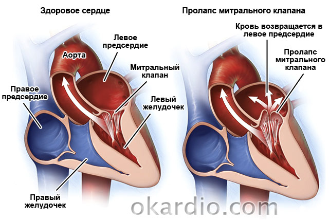
Another common pathology is. It occurs in 2.4% of the population. This is a congenital defect in which the valve leaflets "sink" into the left atrium. In 30% of cases, it is asymptomatic. In the remaining 70% of patients, doctors note shortness of breath, pain in the heart area, accompanied by nausea and a feeling of "lump" in the throat, arrhythmias, fatigue, dizziness, frequent fever up to 37.2–37.4.
Treatment may not be required if the disease is asymptomatic. If the defect is accompanied by arrhythmias or pain in the heart, symptomatic therapy is prescribed. With a strong change in the valve, surgical correction is possible. Since the disease progresses with age, patients need to be examined by a cardiologist 1-2 times a year.
Ebstein anomaly

Ebstein's anomaly is the displacement of the tricuspid valve leaflets into the right ventricle. Symptoms: shortness of breath, paroxysmal tachycardia, fainting, swelling of the veins in the neck, enlargement of the right atrium and the upper part of the right ventricle.
Treatment for asymptomatic course is not carried out. If the signs are pronounced, surgical correction or valve transplantation is done.
congenital heart defects
Congenital anomalies of the structure of the heart include:
- An atrial septal defect is a communication between the right and left atria.
- A ventricular septal defect is an abnormal communication between the right and left ventricles.
- The Eisenmenger complex is a ventricular septal defect located high, the aorta is displaced to the right and connects simultaneously with both ventricles (aortic dextroposition).
- An open ductus arteriosus - the communication between the aorta and the pulmonary artery, which is normally present at the embryonic stage of development, has not been overgrown.
- Tetralogy of Fallot is a combination of four defects: ventricular septal defect, aortic dextroposition, pulmonary artery stenosis, and right ventricular hypertrophy.

Congenital heart defects - signs and treatment:
| Name | Symptoms | Treatment |
|---|---|---|
| Atrial septal defect | With a small defect, signs begin to appear in middle age: after 40 years. This is shortness of breath, weakness, fatigue. Over time, chronic heart failure develops with all the characteristic symptoms. The larger the size of the defect, the sooner the symptoms begin to appear. | Surgical closure of the defect. It is not always carried out. Indications: ineffectiveness of medical treatment of CHF, lag in physical development in children and adolescents, increased blood pressure in the pulmonary circle, arteriovenous discharge. Contraindications: veno-arterial discharge, severe left ventricular failure. |
| Ventricular septal defect | If the defect is less than 1 cm in diameter (or less than half the diameter of the aortic orifice), only shortness of breath during physical exertion of moderate intensity is characteristic. If the defect is larger than the indicated dimensions: shortness of breath with little exertion or at rest, pain in the heart, cough. |
Surgical closure of the defect. |
| Eisenmenger complex | Clinical picture: cyanosis of the skin, shortness of breath, hemoptysis, signs of CHF. | Medication: beta-blockers, endothelin antagonists. Surgery to close a septal defect, correct aortic origin, and replace an aortic valve is possible, but patients often die during surgery. The average life expectancy of the patient is 30 years. |
| Tetralogy of Fallot | Blue tint of mucous membranes and skin, retardation in growth and development (both physical and intellectual), convulsions, low blood pressure, symptoms of CHF. The average life expectancy is 12-15 years. 50% of patients die before the age of 3 years. |
Surgical treatment is indicated for all patients without exception. In early childhood, surgery is performed to create an anastomosis between the subclavian and pulmonary arteries in order to improve blood circulation in the lungs. At the age of 3–7 years, a radical operation can be performed: simultaneous correction of all 4 anomalies. |
| Open ductus arteriosus | A long time proceeds without clinical signs. Over time, shortness of breath and a strong heartbeat, pallor or a blue tint of the skin, and low diastolic blood pressure appear. | Surgical closure of the defect. It is shown to all patients, except for those who have a shunt of blood from right to left. |
Inflammatory diseases
Classification:
- Endocarditis - affects the inner lining of the heart, the valves.
- Myocarditis - muscular membrane.
- Pericarditis - pericardial sac.

They can be caused by microorganisms (bacteria, viruses, fungi), autoimmune processes (eg rheumatism) or toxic substances.
Also, inflammation of the heart can be complications of other diseases:
- tuberculosis (endocarditis, pericarditis);
- syphilis (endocarditis);
- flu, tonsillitis (myocarditis).
Pay attention to this and consult a doctor in time if you suspect flu or sore throat.
Symptoms and treatment of inflammation
| Name | Symptoms | Treatment |
|---|---|---|
| Endocarditis | High temperature (38.5–39.5), increased sweating, rapidly developing valvular defects (detected by echocardiography), heart murmurs, enlarged liver and spleen, increased vascular fragility (hemorrhages under the nails and in the eyes can be seen), thickening of the tips fingers. | Antibacterial therapy for 4-6 weeks, valve transplantation. |
| Myocarditis | It can occur in several ways: attacks of pain in the heart; symptoms of heart failure; or with extrasystoles and supraventricular arrhythmias. An accurate diagnosis can be made on the basis of a blood test for cardiospecific enzymes, troponins, and leukocytes. | Bed rest, diet (No. 10 salt restricted), antibacterial and anti-inflammatory therapy, symptomatic treatment of heart failure or arrhythmias. |
| Pericarditis | Chest pain, shortness of breath, palpitations, weakness, cough without sputum, heaviness in the right hypochondrium. | Non-steroidal anti-inflammatory drugs, antibiotics, in severe cases - subtotal or total pericardectomy (removal of part or all of the pericardial sac). |
Rhythm disturbances
Causes: neurosis, obesity, malnutrition, cervical osteochondrosis, bad habits, intoxication with drugs, alcohol or drugs, coronary artery disease, cardiomyopathy, heart failure, premature ventricular excitation syndromes. The latter are heart diseases in which there are additional pathways for conducting an impulse between the atria and ventricles. You will read about these anomalies in a separate table.
Characteristics of rhythm disturbances:
| Name | Description |
|---|---|
| Sinus tachycardia | Rapid heart rate (90-180 per minute) while maintaining a normal rhythm and a normal pattern of impulse propagation through the heart. |
| Atrial fibrillation (flicker) | Uncontrolled, irregular and frequent (200-700 per minute) atrial contractions. |
| atrial flutter | Rhythmic atrial contractions with a frequency of about 300 per minute. |
| ventricular fibrillation | Chaotic, frequent (200–300 per minute) and incomplete contractions of the ventricles. The lack of full contraction provokes acute circulatory failure and fainting. |
| ventricular flutter | Rhythmic contractions of the ventricles with a frequency of 120–240 per minute. |
| Paroxysmal supraventricular (supraventricular) tachycardia | Attacks of rhythmic palpitations (100-250 per minute) |
| Extrasystole | Spontaneous contractions out of rhythm. |
| Conduction disorders (sinoatrial block, interatrial block, atrioventricular block, bundle branch block) | Deceleration of the rhythm of the whole heart or individual chambers. |

Syndromes of premature excitation of the ventricles:
| WPW syndrome (Wolf-Parkinson-White syndrome) | CLC syndrome (Clerk-Levi-Christesco) |
|---|---|
| Signs: paroxysmal (paroxysmal) supraventricular or ventricular tachycardia (in 67% of patients). Accompanied by a feeling of increased heartbeat, dizziness, sometimes - fainting. | Symptoms: tendency to attacks of supraventricular tachycardia. During them, the patient feels a strong heartbeat, dizziness may occur. |
| Reason: the presence of a bundle of Kent - an abnormal conduction path between the atrium and the ventricle. | Cause: The presence of the James bundle between the atrium and the atrioventricular junction. |
| Both diseases are congenital and quite rare. | |
Treatment of rhythm disorders
It consists in the treatment of the underlying disease, correction of diet and lifestyle. Antiarrhythmic drugs are also prescribed. Radical treatment for severe arrhythmias is the installation of a defibrillator-cardioverter, which will "set" the rhythm of the heart and prevent ventricular or atrial fibrillation. With conduction disorders, pacing is possible.

Treatment of ventricular preexcitation syndromes can be symptomatic (elimination of seizures with medication) or radical (radiofrequency ablation of the abnormal conduction pathway).
Cardiomyopathy
These are myocardial diseases that cause heart failure, not associated with inflammatory processes or pathologies of the coronary arteries.
The most common are hypertrophic and. Hypertrophic is characterized by the growth of the walls of the left ventricle and the interventricular septum, dilated - an increase in the cavity of the left, and sometimes the right ventricles. The first is diagnosed in 0.2% of the population. It occurs in athletes and can cause sudden cardiac death. But in this case, it is necessary to conduct a thorough differential diagnosis between hypertrophic cardiomyopathy and non-pathological heart enlargement in athletes.

- congenital aortic stenosis;
- coarctation of the aorta;
- transposition of the great vessels;
- total anomaly of the pulmonary veins;
- coarctation of the aorta and PDA.
Ecology
Changes at the genetic level can be caused by exposure to 3 main mutagens:
Genetics
The most common causes of congenital heart disease include point gene changes, as well as chromosomal mutations, manifested by deletion or duplication of DNA segments. Common mutations include trisomies 21,13,18.
There is an association between specific genes and certain malformations. For example, it has been found that a mutation in myocardial muscle protein leads to the development of atrial septal defects.
Pathogenesis
The development of heart defects is based on 2 mechanisms:
| Violation of cardiac hemodynamics |
|
| Violation of systemic hemodynamics | Leads to the development of systemic hypoxia. |

Types of heart defects
Depending on the mechanism of formation of pathology, it is divided into two types:
| birth defects |
|
| Acquired vices | They can form at any age. The main reasons that can lead to the development of pathology are: rheumatism, hypertension, syphilis, coronary disease, atherosclerotic vascular lesions, cardiosclerosis, as well as severe injuries of the chest (myocardium). |
There is also another classification:
The classification depending on the localization is as follows:
- valve defects;
- defects of the interventricular septum, as well as the interatrial septum.

You can also highlight:
Also, the degree of defect, which can be from I to IV, should be indicated in the outpatient card.
Clinical picture
Symptoms depend on the severity of the lesions. In the bulk, signs are detected within a few years after birth, however, there are cases when the pathology is not detected throughout life.
As a rule, the idea of the presence of violations can be suggested by noises heard during auscultation. At the same time, not all murmurs are due to heart defects.
Clinically, heart defects are described by 4 syndromes:
- cardiac: chest pain, tachycardia, dizziness, vascular pulsation;
- heart failure syndrome;
- chronic systemic hypoxia syndrome: non-compliance with developmental standards, symptoms of drumsticks, watch glasses;
- respiratory disorders syndrome is characteristic of congenital heart defects with enrichment of the pulmonary circulation.
Frequently asked Questions
How many live with congenital or acquired pathology?
It should be noted right away that not all diagnoses with the wording “heart disease” should cause panic. Some of them are quite harmless and do not pose a risk to life if the simplest recommendations of a specialist are followed.
It is necessary to adhere to proper nutrition, avoid excessive physical exertion, stress. If these conditions are met, it is possible to live to a respectable age without experiencing health problems caused by PS.
Moreover, there are cases when people with undiagnosed pathology lived for a long time and did not show any symptoms.
However, in cases where the disease is diagnosed, it is necessary to conduct regular examinations and follow preventive measures. Even a minor defect that does not bring discomfort, sooner or later, under the influence of various factors, can become dangerous. Therefore, it is impossible to say how many people live with heart disease without surgery - each patient has his own history of the development of the disease.
Heart defects can be manifested by damage to the heart muscle of varying severity, disruption of the valves, the result of which in most cases is heart failure.
Violation of the mechanisms of the heart leads to a deterioration in the circulation of the body as a whole. This is accompanied by an increased likelihood of developing a heart attack or stroke.
Advanced forms of pathology are accompanied by a high risk of death. With defects that are accompanied by a risk to life, surgical intervention is recommended.
There are a number of signs, in the event of which it is necessary to urgently consult a specialist:
- dyspnea;
- swelling of the lower extremities;
- blueing of the skin;
- rapid increase in body weight;
- constant dizziness;
- fainting states;
- body temperature lability;
- nausea;
- itching in the limbs.

Is it dangerous?
Even a congenital form of heart disease may not manifest itself immediately. Up to the third year of life, any symptoms of the development of pathology may be absent. Among the signs that may indicate a violation include: a lag in physical development, shortness of breath during physical exertion, pallor or cyanosis of the skin.
The clinical picture of defects that occur against the background of blue skin, includes the following symptoms: anxiety, shortness of breath, high probability of fainting. Seizures usually develop suddenly. This condition is typical for children under 2 years of age.
The defects in which pallor of the skin develops are accompanied by a developmental delay that affects the lower half of the body, the occurrence of headaches and dizziness, pain in the chest, abdomen, and lower extremities.
Is it hereditary
It is very important for future parents who have this pathology in their medical history to know whether congenital heart disease is inherited.
The tendency to form pathology associated with structural and functional disorders of the myocardium can be transmitted from parent to child.
Today, medicine has the capabilities that allow you to determine the pathology at an early stage, which makes it possible to immediately respond to the changes taking place.
A congenital anomaly in the development of organs occurs during the period of intrauterine development of the fetus. Recently, more and more doctors are faced in their practice with various kinds of heart defects in newborns. However, one of the rarest types of congenital pathology in the fetus is Cantrell's pentad syndrome. Description...
The upper part of the heart consists of the right and left atria, separated in the middle by a longitudinal septum without any gaps. An atrial septal defect is a congenital heart defect characterized by a hole in the septum that facilitates communication between the right and left atrium. Primary...
The term "heart defects" in medicine means a whole range of anomalies in the structure of the heart muscle, which in one way or another affect the functioning of the organ. In this case, the following can be affected: the walls of the heart chambers themselves; valves involved in the differentiation of chambers; blood vessels that take blood away from the heart and lead to it ...
The Eisenmenger complex in medicine today is understood as a complex cardiac pathology, which involves the interventricular septum, aorta, and right ventricle. At the same time, there is a defect in the interventricular septum, the right ventricle has pronounced signs of hypertrophy, and the aorta ...
Various types of heart defects are a very common cause of early childhood mortality. According to statistics, the susceptibility of newborns to pathologies of this group is 5%. At the same time, in 2% of cases, children cannot be saved, since the defects give a clinical picture that is incompatible with ...
Heart disease is a pathology of the valves, which leads to disruption of the organs. The problem develops during fetal development or may be the result of previous diseases. Defects often do not manifest themselves for years, and a person learns about them during a routine examination. The disease requires treatment, as it can become a cause.
What is heart disease and why is it dangerous
Defects are called anomalies in the structure and dysfunction of the heart valves, stenosis of the openings they close, defects in the walls and partitions. The pathological process affects one valve or several at once.
In connection with these violations, the valve does not fulfill its purpose, that is, it does not completely block the hole located under it. As a result, during the contraction, the blood returns to the cardiac chamber from which it has just left. This causes congestion in the chamber above and overloads the heart.
The lower chambers fill with blood during relaxation. The same condition is observed with vascular stenosis.
The constant overflow of the chambers disrupts the blood flow. Due to its plasticity, the heart adapts to this state. To cope with the load, the organ increases the thickness of the myocardium and the volume of the chambers.
This condition is called a compensated defect.
The heart muscle in such conditions needs more nutrition. Therefore, there is a need for increased blood flow. If the blood supply does not increase, then the myocardium weakens, and the stage of decompensation develops.
There are various vices. In general, there are about a hundred species.
Each poses a certain danger to human health. The disease almost always requires surgical intervention, but each case differs in individual characteristics.
Sometimes surgery is used to eliminate. There are types of pathology that are non-fatal, so the operation is postponed for several years.
The risk of complications during surgery is especially high if the patient is a newborn. During the period for which the surgical procedures were postponed, the patient must take medication.
Therapeutic procedures can ease the work of the heart and improve well-being.
Kinds
Heart defects are divided into two large groups. They can be:
- Congenital. Their formation occurs during fetal development. During the laying of organs and systems, some kind of failure may occur under the influence of external and internal factors. The external ones include an unfavorable ecological situation, viral and infectious diseases during pregnancy, the use of certain drugs, an unhealthy lifestyle of a pregnant woman. Deviations can be associated with hormonal changes, hereditary predisposition.
- Acquired. They develop at any age under the influence of infectious and autoimmune diseases. In most cases, the defect appears with syphilis, hypertension, ischemia, rheumatism, atherosclerosis.
There are also vices:
- simple, when changes occur in one valve;
- complex, in which two or more violations are combined. For example, stenosis is combined with valvular insufficiency;
- combined. They are presented as a complex combination of disorders affecting several valves or orifices.
Given the place in which they are located, the vices are:
- mitral, aortic, tricuspid, pulmonary valve;
- partitions between the ventricles and the atria.
To fully characterize pathologies, they are divided into:
- White. With them, venous blood does not mix with arterial, and the tissues do not suffer from a lack of oxygen.
- Blue. Venous blood is mixed and thrown into the arterial bed. This is accompanied by the expulsion of insufficiently oxygenated blood, the patient suffers from manifestations of heart failure.
There are also four degrees of development of defects, which are distinguished depending on the characteristics of circulatory disorders.
Acquired vices
Pathology often ends with disability or death at a young age. In most cases, the defects are caused by rheumatism. The cause of development can also be atherosclerotic changes, infection with syphilis.
The structure of the heart is disturbed as a result of prolonged sepsis, trauma and tumors.
 Most often, valvular defects develop. People in their 30s usually have mitral or tricuspid regurgitation. Aortic insufficiency as a result of syphilis occurs after 50 years. Defects in atherosclerosis affect people after 60 years.
Valvular insufficiency is characterized by a partial return of the blood ejected during contraction, overflowing the heart and causing congestion.
If the heart hole narrows, the same disorders occur. Due to the narrow opening, blood passes through the vessels and the next chamber with great difficulty, causing overflow and distention.
If rheumatism develops rapidly, as a result of complications, the patient becomes disabled.
With syphilis, chronic sepsis, severe atherosclerosis, the valves become denser, and their mobility is impaired, because of this, the valves do not close completely and insufficiency develops. Part of the blood at the exit from the left ventricle returns back, which is accompanied by its expansion and thickening of the muscles.
In a severe form of circulatory failure, pulmonary edema develops or.
At the same time, the patient's skin turns pale, the vessels of the neck pulsate, and the head sways to the beat of the pulsation. Insufficient oxygen supply causes headaches and dizziness, discomfort in the heart area.
Blood pressure indicators are also subject to changes: systolic values increase, and diastolic values decrease.
If the insufficiency struck the tricuspid valve, then other deviations most often join it. The problem is often formed in combination with mitral stenosis.
Swelling and puffiness of the face, blue skin to the waist are considered to be special manifestations of the problem.
What will be the prognosis for such defects depends on the timeliness and correctness of the treatment of heart lesions. Well-being usually worsens during stress, during pregnancy and physical activity.
birth defects
Heart disease is a pathology that can occur in the prenatal period. They develop in violation of the process of laying organs in the embryonic period. The exact causes of the pathology have not yet been determined. But the presence of infectious diseases in women, such as influenza, rubella, syphilis, viral hepatitis, has a certain effect.
Improper nutrition and vitamin deficiency, the negative effects of radiation can also lead to developmental failures. Most often, the ductus arteriosus, interventricular and atrial septum are not closed. In rare cases, narrowing of the pulmonary artery and isthmus of the aorta.
A small percentage of children develop an isolated defect.
All these pathologies are accompanied by complex combined anatomical changes in the structure of the heart.
If a child was born with a defect, then his skin turns blue, his fingers thicken at the ends and become similar to drumsticks.
The reasons for these changes are insufficient oxygen supply to the tissues. The mortality rate from this pathology used to be quite high. Today, the development of cardiac surgery allows you to save the lives of children.
The ductus arteriosus is responsible for connecting the artery and the aorta. In the intrauterine period, it remains open, before childbirth, this duct closes. But sometimes this does not happen, because of which the blood overflows from the ventricle to the ventricle, causing them to increase in size.
If the hole is large, then the clinical manifestations will be pronounced. For treatment, surgical techniques are used, consisting in stitching the duct and its complete closure.
Violation in the structure of the interventricular septum is characterized by the presence of an open two-centimeter hole.
Under the influence of high pressure of the left ventricle, the blood passes to the right side, causing expansion of the right ventricle and congestive processes in the lungs.
In connection with compensatory mechanisms, an increase in the left ventricle occurs. If the patient does not feel any discomfort, then the problem can be detected by auscultation, which allows you to detect heart murmurs.
To eliminate the problem, surgery is performed, during which the hole is closed with synthetic materials.
Septal defects between the atria occur in 20% of cases. They often make up and.
In the area between the atria, there is an oval foramen, which closes in childhood. But it doesn't always close.
On the left side, the hole covers the valve leaf and presses it tightly under the influence of high pressure in this area of the heart. But if mitral stenosis develops, there is an increase in pressure in the right side and there is a penetration of blood from the right side into the left.
To eliminate the problem, surgical methods of treatment are used. If there is a small defect, the hole is sewn together. If it is large, then grafts and prosthetic materials are used for closure.
Such anomalies in the development of the heart are dangerous with the possibility of thromboembolism.
An X-ray with a contrast agent is used to make the diagnosis. If the substance is injected into the cardiac chamber, then it is distributed by open ducts.
It is most difficult to carry out treatment if one patient has four or more defects combined at once. This phenomenon is called Fallot's tetrad.
To eliminate congenital defects and prevent the development of their complications, treatment should be carried out at the initial stages, avoiding decompensation.
Patients should be under dispensary observation for protection against infectious diseases, control of diet and level of physical activity.
Life expectancy with defects
How long they live with heart disease is of interest to all patients. Calculating the exact time is quite difficult. Many factors influence the future prognosis. To predict the consequences, the type of defect, the severity of the development of the pathology, the age of the patient, the general health of the patient, the presence of concomitant pathologies and other features are taken into account.
The most difficult organism tolerates combined defects. No less negatively affect the health and pathology of neighboring organs. The heart is closely connected with the lungs, liver, and circulatory system.
Therefore, in the presence of pathological processes in these organs, one cannot count on a favorable outcome. It is also important to take into account what kind of lifestyle a person leads, his diet, working and living conditions, regular walks in the fresh air.
The most difficult thing is to predict the consequences of congenital malformations. Very often, children do not manage to live for several years. But with the help of advances in cardiac surgery, the death rate has been reduced. Many people live with such a diagnosis for decades, if they treat their health correctly, take medications and follow all the doctor's recommendations.
Each patient should undergo an examination every six months to assess the condition of the heart. If violations are detected, then surgical treatment will be urgently performed.
Heart defects - a group of diseases characterized by a violation of the anatomical structure of the heart valves or partitions. They are usually manifested by hemodynamic disturbances.
These diseases occur in both adults and children and may be associated with a number of factors. To understand what a vice is, you need to know not only the causes of this disease, but also how it affects the overall functioning of the body.
Classification

The structure of the heart
To understand what heart defects are, you need to know their classification. To begin with, it should be said that there are congenital and acquired defects.
Depending on the location of the lesion, there are the following types of defects:
- valve pathology.
- Partition pathologies.
According to the number of affected structures, the following types of heart defects are distinguished:
- Simple. One valve failure.
- Complex. Damage to multiple valves.
- Combined. The combination of insufficiency and stenosis in one valve.
The classification of heart defects also takes into account the level of general hypoxia of the body. Depending on this factor, the following types are distinguished:
- White. Sufficient supply of tissues and organs with oxygen. Cyanosis is not formed.
- Blue. Early manifestation of hypoxia, the formation of central cyanosis.
Also, for the correct classification of congenital forms, the Marder table is used:
| CHANGES IN HEMODYNAMICS | WHITE | BLUE |
| not violated | Right-sided heart, aortic malposition, minor ventricular septal defect (VSD) | - |
| Overfilling the small circle | VSD, patent ductus arteriosus, abnormal pulmonary venous drainage | Transposition of the main vessels, ventricular underdevelopment, common truncus arteriosus |
| Impoverishment of the small circle | Stenosis of the pulmonary artery | Tetralogy of Fallot, false common truncus arteriosus, Ebstein's disease |
| Violation of blood flow in a large circle | Stenosis of the mouth and coarctation of the aorta | - |
Causes

The etiology of congenital forms is represented by various chromosomal disorders. Environmental factors also play a role. In the event that the mother had rubella during pregnancy, the child has the following three syndromes:
- Cataract.
- Deafness.
- Congenital anomalies of the heart. Most often it can be Fallot's tetrad (a combined congenital defect, which includes stenosis of the outflow tract of the right ventricle, aortic displacement, right ventricular hypertrophy).
- In addition to rubella, the following infectious agents have an impact on the embryonic development of the cardiovascular system:
- Herpes simplex virus.
- adenovirus.
- Cytomegalovirus.
- Mycoplasma.
- Toxoplasma.
- Pale treponema.
- Listeria.
The etiology of congenital malformations is quite wide. It includes the use of alcohol and drugs by a pregnant woman. The teratogenic effect on the fetus of amphetamines has been proven.
Despite the fact that defects are not inherited, the presence of a family history of cardiovascular pathologies increases the risk of valvular heart disease in children. Also, some experts talk about the possible development of a congenital defect in the next generations with a closely related marriage.
The most common cause is acute rheumatic fever. In addition, the etiology includes the following diseases:
- Atherosclerosis.
- Syphilis.
- Autoimmune diseases.
- Heart injury.
Main manifestations
The pathophysiology of congenital heart defects to a greater extent lies in the disruption of normal blood circulation. Most often this contributes to:
- Increased resistance to blood flow, leading to hypertrophy of the corresponding cardiac section.
- Creation of a vicious communication between both circles of blood circulation.
Very often, both of these pathogenetic mechanisms can be combined. In this regard, the following signs of heart defects can be distinguished:
- Dyspnea.
- pathological noises.
- Fainting states.
- Cyanosis.
- Palpitation.
- Rhythm disturbance.
- Peripheral edema in the legs.
- Ascites (accumulation of fluid in the abdominal cavity).
- Hydrothorax (accumulation of fluid in the pleural cavity).
It is worth noting that compensated defects are most often asymptomatic and are detected during a routine examination. The main symptoms of heart defects appear when the body can no longer cope with hemodynamic disorders on its own.
Diagnostics
 Based only on the symptoms identified during the examination, the diagnosis of "heart disease" is impossible. To do this, the doctor needs to conduct a number of physical and instrumental methods. Directly at the first visit to a specialist, propaedeutics of internal diseases plays an important role, in particular, auscultation and palpation examination.
To understand how to determine heart disease, it is important to know the basic instrumental methods used in the diagnosis of such diseases. The following studies are applied:
- Electrocardiography. An electrocardiogram will help detect changes in the work of the heart. ECG for heart defects does not play a big role and most often helps to identify the consequences of the disease.
- Echocardiography. This is an ultrasound diagnostics designed for a thorough study of the functional and morphological features of the heart.
- Phonocardiography is a technique that allows you to display heart sounds in a graphical form.
- X-ray. Allows you to identify the configuration of the heart, as well as to detect the transposition of blood vessels.
Modern pediatrics assigns a special place in the definition of congenital anomalies to prenatal detection. Such a diagnosis of heart defects is carried out in several stages. The initial stage lies on the shoulders of a regular gynecologist, and after suspicion of the presence of changes, the pregnant woman is sent to a specialist in fetal echocardiography.
Therapeutic measures
The main method of therapy for most heart defects is surgery. Nevertheless, in order to reduce the intensity of the progression of the disease, as well as normalize blood circulation, the following groups of drugs are used:
- Anticoagulants. Their use is explained by the need to reduce the risk of thrombosis.
- Diuretics. Diuretics are needed to relieve edematous syndrome, as well as to compensate for heart failure.
- β-blockers. Necessary to reduce the load on the heart and normalize blood pressure.
- ACE inhibitors. Like previous drugs, they are needed to combat arterial hypertension.
- Antiarrhythmic drugs. They are used for defects during which there is a violation of the heart rhythm.
- Vasodilators. Reduce stress on the heart.
 About how to treat heart disease in each case, only a doctor can answer, having previously made a diagnosis. It is extremely important to follow all medical recommendations and use drugs strictly in the indicated dosage.
Methods of surgical treatment of heart disease in adults and children are quite diverse. Treatment is used for both acquired and congenital malformations. Indications for surgical intervention directly depend on the degree of hemodynamic disturbance. In congenital forms and in the presence of an obstruction to blood flow, the following interventions are performed to eliminate it:
- Dissection of the valvular constriction in the pulmonary trunk.
- Excision of narrowed areas in the aorta.
In the event that the defect is accompanied by overflow of the small circle, it is necessary to eliminate the pathological discharge of blood. This is achieved through the following activities:
- Ligation of the open aortic duct.
- Sewing up an existing atrial or interventricular septal defect.
In addition to the above, in congenital forms, the following operations of heart defects are used:
- valve plastic.
- Prosthetics.
- Reconstruction.
Reconstruction is used for complex pathologies, which are accompanied not only by a violation of the valvular apparatus, but also by the incorrect location of large vessels.
For a long time, it was believed that surgical treatment of congenital heart defects should be carried out at the age of five or even later. However, the level of medicine currently allows for surgical correction in the first years of life.
With acquired pathologies of the valvular apparatus, the main surgical intervention is prosthetics. The patient is replaced by an artificial valve in place of the damaged one.
With acquired stenoses, in order to eliminate the existing obstacle, valve plastic is performed.
Complications
To understand why heart disease is dangerous, you need to know what consequences can occur. The main complications are associated with the formation of circulatory failure. The following types are distinguished:
- Left ventricular failure.
- Right ventricular failure.
One of the main signs of severe left ventricular failure is shortness of breath, accompanied by the release of bloody frothy sputum. Its color is due to the presence of special cells. Cells of heart defects are represented by alveolar macrophages containing hemosiderin. The presence of these cells is observed in pulmonary infarction or stagnation in the pulmonary circulation. And it is precisely because of them that sputum with these pathologies has a “rusty” color.
Nursing Process
The nursing process for heart defects plays an important role in improving the patient's quality of life. This process has the following goals:
- Improving the quality of life of the patient to an acceptable level.
- Minimization of emerging problems.
- Help the patient and his family in adapting to the disease.
- Support for the patient's basic needs or their full recovery.
With heart defects, this process is carried out in several stages. At the first stage, the nurse must determine the basic needs of the patient. The second stage consists in making a nursing diagnosis and identifying the main problems of the patient, among which the following should be highlighted:
- Physiological. This group includes all complications of the main diagnosis.
- Psychological. Anxiety about changes in the quality of life, as well as the fear of worsening the prognosis of the disease. It also includes a lack of knowledge and skills in general health care.
- Security issues. Attention should be paid to the increased risk of infection, misuse of prescribed medications, and the risk of falls if fainting occurs.
 The third stage is to determine the goals that the nurse should set for herself in order to alleviate the general condition of the patient. The fourth stage includes the implementation of all nursing interventions that must be carried out according to the protocol for the treatment and diagnosis of the defect. The last stage - an assessment of how effectively nursing care was carried out, is carried out not only by the nurse, but also by her immediate superiors, as well as by the patient himself.
Preventive actions
Prevention of heart defects is an extremely important set of measures to reduce the risk of violations in the operation of the valvular apparatus of the heart. It is worth noting that in order to prevent congenital malformations, the expectant mother must follow the following recommendations:
- Timely registration.
- Regular visits to the gynecologist at the scheduled time for a preventive examination.
- Proper nutrition.
- Exclusion of bad habits.
- Correction of concomitant diseases.
- If not vaccinated against rubella virus, vaccinate approximately 6 months before planned conception.
- Prophylactic intake of folic acid.
The main cause of acquired heart defects is rheumatism. That is why one of the main preventive measures is to prevent the development of acute rheumatic fever.
- Leading a healthy lifestyle.
- Proper nutrition.
- Timely treatment of exacerbations of chronic pathologies.
- Sanitation of infectious foci.
After treatment of adults and adolescents for rheumatism, bicillin prophylaxis is recommended.
Also, in order to reduce the manifestations of the rheumatic process, as well as to prevent possible consequences, it is recommended to massage with heart defects. There are the following indications for massage:
- inactive stage of the disease.
- The period of reduction of the main manifestations.
- Formed circulatory failure.
During the period of active inflammatory changes in the cardiac membranes, the use of massage is not allowed.