What organs are formed from the germ layers. germ layer theory. Derivatives of the germ layers. The history of the development of the theory of germ layers
Germ layers is a basic term in embryology. They designate the layers of the fetal body at an early stage. In most cases, these layers are epithelial in nature.
The germ layers are usually classified into three types:
Ectoderm - the outer sheet, which is also called the epiblast or skin-sensitive layer;
Endoderm is the inner layer of cells. It may also be called a hypoblast or entero-glandular sheet;
Middle layer (mesoderm or mesoblast).
Germinal sheets (depending on their location, they are characterized by certain cell features. Thus, the outer layer of the embryo consists of light and tall cells, which are similar in structure to cylindrical epithelium. The inner leaf consists in most cases of large cells that are filled with specific yolk plates. They have a flattened appearance, which makes them look like
The mesoderm at the first stage consists of spindle-shaped and stellate cells. They later form the epithelial layer. I must say that many researchers believe that the mesoderm is the middle germ layers, which are not an independent layer of cells.
The germ layers at first have the form of a hollow formation, which is called the blastodermal vesicle. At one of its poles, a group of cells gathers, which is called a cell mass. It gives rise to the primary intestine (endoderm).
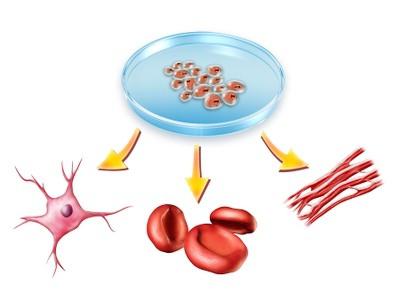
It should be said that different organs are formed from embryonic sheets. Thus, the nervous system arises from the ectoderm, the digestive tube originates from the endoderm, and the skeleton and muscles originate from the mesoderm.
It should also be noted that special embryonic membranes are formed during embryogenesis. They are temporary, do not participate in the formation of organs and exist only during embryonic development. Each class has certain features in the formation and structure of these shells.

With the development of embryology, they began to determine the similarity of embryos, which was first described by K.M. Baer in 1828. A little later, Charles Darwin identified the main reason for the similarity of embryos of all organisms - their common origin. Severov, on the other hand, argued that the common signs of embryos are associated with evolution, which proceeds in most cases through anabolism.
When comparing the main stages of development of embryos of different classes and species of animals, certain features were found, which made it possible to formulate the law of embryonic similarity. The main provisions of this law was that the embryos of organisms of the same type in the early stages of their development are very similar. Subsequently, the embryo is characterized by more and more individual features that indicate its belonging to the corresponding genus and species. At the same time, the embryos of representatives of the same type are increasingly separated from each other, and their primary similarity is no longer traced.
germ layers(lat. embryonic folia), germ layers, layers of the body of the embryo of multicellular animals, formed during gastrulation and giving rise to various organs and tissues. In most organisms, three germ layers are formed: the outer one is the ectoderm, the inner one is the endoderm and the middle mesoderm.
Derivatives of the ectoderm perform mainly integumentary and sensitive functions, derivatives of the endoderm - the functions of nutrition and respiration, and derivatives of the mesoderm - connections between parts of the embryo, motor, support and trophic functions.
The same germ layer in representatives of different classes of vertebrates has the same properties, i.e. germ layers are homologous formations and their presence confirms the position of the unity of the origin of the animal world. Germ layers are formed in embryos of all major classes of vertebrates, i.e. are universally distributed.
The germ layer is a layer of cells that occupies a certain position. But it cannot be considered only from topographic positions. The germ layer is a collection of cells that have certain developmental tendencies. A clearly defined, albeit rather wide, range of developmental potentials is finally determined (determined) by the end of gastrulation. Thus, each germ layer develops in a given direction, takes part in the emergence of the rudiments of certain organs. Throughout the animal kingdom, individual organs and tissues originate from the same germ layer. From the ectoderm, the neural tube and integumentary epithelium are formed, from the endoderm - the intestinal epithelium, from the mesoderm - muscle and connective tissue, the epithelium of the kidneys, gonads, and serous cavities. From the mesoderm and the cranial part of the ectoderm cells are evicted, which fill the space between the sheets and form the mesenchyme. Mesenchymal cells form syncytium: they are connected to each other by cytoplasmic processes. The mesenchyme forms the connective tissue. Each individual germ layer is not an autonomous formation, it is part of the whole. The germ layers are able to differentiate only by interacting with each other and being under the influence of the integrating influences of the embryo as a whole. A good illustration of such interaction and mutual influence are experiments on early gastrulae of amphibians, according to which the cellular material of the ecto-, ento- and mesoderm can be forced to radically change the path of its development, to participate in the formation of organs that are completely uncharacteristic of this leaf. This suggests that, at the beginning of gastrulation, the fate of the cellular material of each germ layer, strictly speaking, is not yet predetermined. The development and differentiation of each leaf, their organogenetic specificity, is due to the mutual influence of the parts of the whole embryo and is possible only with normal integration.
62. Histo- and organogenesis. The process of neurulation. Axial organs and their formation. mesoderm differentiation. Derivative organs of vertebrate embryos.
Histogenesis(from other Greek ἱστός - tissue + γένεσις - education, development) - a set of processes leading to the formation and restoration of tissues in the course of individual development (ontogenesis). One or another germ layer is involved in the formation of a certain type of tissue. For example, muscle tissue develops from mesoderm, nervous tissue from ectoderm, etc. In some cases, tissues of the same type may have a different origin, for example, the epithelium of the skin is ectodermal, and the absorbent intestinal epithelium is endodermal in origin.
Organogenesis- the last stage of embryonic individual development, which is preceded by fertilization, crushing, blastulation and gastrulation.
In organogenesis, neurulation, histogenesis and organogenesis.
In the process of neurulation, a neurula is formed, in which the mesoderm is laid, consisting of three germ layers (the third layer of the mesoderm splits into segmented paired structures - somites) and the axial complex of organs - the neural tube, chord and intestine. The cells of the axial complex of organs mutually influence each other. This mutual influence is called embryonic induction.
In the process of histogenesis, body tissues are formed. From the ectoderm, nervous tissue and the epidermis of the skin with skin glands are formed, from which the nervous system, sensory organs and epidermis subsequently develop. From the endoderm, a notochord and epithelial tissue are formed, from which mucous membranes, lungs, capillaries and glands (except for the genital and skin ones) are subsequently formed. The mesoderm produces muscle and connective tissue. ODS, blood, heart, kidneys and gonads are formed from muscle tissue.
Neurulation- the formation of the neural plate and its closure into the neural tube in the process of embryonic development of chordates.
Neurulation is one of the key stages of ontogeny. An embryo at the stage of neurulation is called a neurula.
The development of the neural tube in the anterior-posterior direction is controlled by special substances - morphogens (they determine which of the ends will become the brain), and the genetic information about this is embedded in the so-called homeotic or homeotic genes.
For example, the morphogen retinoic acid, with an increase in its concentration, is able to turn rhombomeres (segments of the neural tube of the posterior part of the brain) of one type into another.
Neurulation in lancelets is the growth of ridges from the ectoderm over a layer of cells that becomes the neural plate.
Neurulation in the stratified epithelium - the cells of both layers descend under the ectoderm mixed, and diverge centrifugally, forming a neural tube.
Neurulation in a single-layered epithelium:
Schizocoelous type (in teleosts) - similar to stratified epithelial neurulation, except that the cells of one layer descend.
In birds and mammals, the neural plate invaginates inward and closes into the neural tube.
In birds and mammals, during neurulation, protruding parts of the neural plate called neural folds, are closed along the entire length of the neural tube unevenly.
Usually, the middle of the neural tube closes first, and then the closure goes to both ends, leaving as a result two open sections - the anterior and posterior neuropores.
In humans, closure of the neural tube is more complex. The spinal section closes first, from the thoracic to the lumbar, the second - the area from the forehead to the crown, the third - the front, goes in one direction, to the neurocranium, the fourth - the area from the back of the head to the end of the cervical, the last, fifth - the sacral section, also goes to one direction, away from the coccyx.
When the second section is not closed, a fatal congenital defect is found - anencephaly. The fetus does not develop a brain.
When the fifth section is not closed, a congenital defect that can be corrected is found - spina bifida, or Spinabifida. Depending on the severity, spina bifida is divided into several subtypes.
During neurulation, the neural tube is formed.
In cross section, immediately after formation, three layers can be distinguished in it, from the inside to the outside:
Ependymal - pseudo-stratified layer containing rudimentary cells.
The mantle zone contains migrating, proliferating cells that emerge from the ependymal layer.
The outer marginal zone is the layer where nerve fibers are formed.
There are 4 axial body: notochord, neural tube, intestinal tube and mesoderm.
Regardless of the animal species, those cells that migrate through the region of the dorsal lip of the blastopore are subsequently transformed into a notochord, and through the region of the lateral (lateral) lips of the blastopore into the third germ layer - the mesoderm. In higher chordates (birds and mammals), due to the immigration of germinal shield cells, the blastopore is not formed during gastrulation. Cells that migrated through the dorsal lip of the blastopore form a notochord, a dense cell strand located along the midline of the embryo between the ectoderm and endoderm. Under its influence, the neural tube begins to form in the outer germinal layer, and only lastly does the endoderm form the intestinal tube.
Differentiation (lat. differens. difference) of the mesoderm begins at the end of the 3rd week of development. The mesenchyme arises from the mesoderm.
The dorsal part of the mesoderm, which is located on the sides of the chord, is divided into body segments - somites, from which bones and cartilage, striated skeletal muscles and skin develop (Fig. 134).
From the ventral non-segmented part of the mesoderm - with the planchnotome, two plates are formed: the splanchnopleura and the somatopleura, from which the mesothelium of the serous membranes develops, and the space between them turns into body cavities, the digestive tube, blood cells, smooth muscle tissue, blood and lymphatic vessels, connective tissue, cardiac striated muscle tissue, adrenal cortex and epithelium sex glands.
Derivatives of the germ layers. The ectoderm gives rise to the outer integument, the central nervous system, and the final section of the alimentary canal. From the endoderm, the notochord, the middle section of the digestive tube and the respiratory system, are formed. From the mesoderm, the musculoskeletal, cardiovascular and genitourinary systems are formed.
Establish a correspondence between the structure of the human body and the germ layer from which it was formed.
Write down the numbers in response, arranging them in the order corresponding to the letters:
| A | B | AT | G | D |
Explanation.
The most important ectodermal derivatives are the neural tube, the neural crest, and all the nerve cells formed from them. The sense organs that transmit information about visual, sound, olfactory and other stimuli to the nervous system also develop from ectodermal anlages. For example, the retina of the eye is formed as an outgrowth of the brain and is therefore a derivative of the neural tube, while olfactory cells differentiate directly from the ectodermal epithelium of the nasal cavity. Pain receptors are of ectodermal origin.
Ectoderm: pain receptors, hairline, nail plates. Mesoderm: lymph and blood, adipose tissue.
Answer: 11221.
Answer: 11221
Source: Unified State Examination in Biology 05/30/2013. main wave. Siberia. Option 2.
Sadi 11.06.2017 13:49
In the answer to this task it is written that the lungs are formed from the mesoderm, and in Task 8 No. 13837 it is said that from the endoderm.
Natalya Evgenievna Bashtannik
Please note that the epithelium of the lungs is the endoderm.
The rudiment of a particular organ is initially formed from a specific germ layer, but then the organ becomes more complex, and as a result, two or three germ layers take part in its formation.
The lung is not only an epithelium, it is also bronchioles, and connecting films ... all this is formed from the mesenchyme, and unfortunately, this knowledge is not considered by the compilers in the Unified State Examination :(
The space between the developing bronchi is filled with intermediate mesenchyme. The mesenchyme, which is a loose tissue tightly covering the developing endodermal tubular formations, begins to differentiate at the root of the lungs in the third month. From here, differentiation continues in the peripheral direction with separate branches of the bronchi. First, the cartilaginous rings of both main bronchi appear, and the cartilaginous plates of the remaining bronchi gradually differentiate. Approximately at the same time, muscle cells and the first collagen fibers of the connective tissue are formed. From the mesodermal material, interlobular and intersegmental septal mesenchyme and subserous connective tissue of the lung film arise. Elastic fibers begin to appear in the fourth month. Their main development occurs, however, as well as the development of cartilaginous plates in the walls of the bronchi, only in the second half of intrauterine development.
"MORDOVA STATE UNIVERSITY named after A.I. N. P. OGAREVA»
Department of Biology
Department of Genetics
on the topic: germ layers
Completed by: 3rd year student
specialty "Biology"
Introduction
1. The structure of the germ layers
2. History of the development of the theory of germ layers
3. Formation of germ layers
4. Origin and evolutionary significance of the germ layers
5. Provisions of the theory of germ layers and objections to this theory
Conclusion
Literature
Introduction
Along with the possibility of interpreting the germ layers from the point of view of their phylogenetic significance, it is important to establish the role they play in individual development. The germ layers are the first organized groups of cells in the embryo, which are clearly distinguished from each other by their features and relationships. The fact that these ratios are basically the same in all vertebrate embryos strongly suggests a common origin and similar heredity in the various members of this vast group of animals.
It can be thought that in these germ layers, for the first time, differences of different classes begin to be created above the general plan of the body structure, characteristic of all vertebrates.
The formation of germ layers ends the period when the main process of development is only an increase in the number of cells, and the period of differentiation and specialization of cells begins. Differentiation occurs in the germ layers before we can see signs of it with any of our microscopic methods. In a leaf that has a completely uniform appearance, localized groups of cells constantly arise with different potentialities for further development.
Various structures arise from the germ layer. At the same time, no visible changes are imperceptible in the germ layer, due to which they arise. Recent experimental studies indicate how early this invisible differentiation precedes the visible morphological localization of cell groups, which we easily recognize as the rudiment of the definitive organ.
1. The structure of the germ layers
The germ layers consist of cellular materials that are used for the development of various organs and tissues. In their structure, the cells of various germ layers differ from each other; endoderm cells are always larger and less regular than ectodermal cells. The endoderm is distinguished by the properties of the future bookmark, which has trophic significance. The ectoderm remains on the surface and initially has a protective value. Unlike the endoderm, it consists of regularly arranged cells of a more uniform shape. Gastrulation leads to a noticeable difference between the outer and inner layers and the germinal material becomes heterogeneous. The process that leads to the appearance of differences in an initially homogeneous material is called differentiation.
Primary organizers or inductors play an important role in the differentiation of cellular material. Inductors are chemicals that are released by groups of cells and affect other groups of cells, changing their developmental path. As a result of differentiation of the germ layers, various organs and tissues are formed. In the study of these processes in different animals, it was found that the fate of each germ layer in all multicellular organisms is, as a rule, the same.
Thus, the epithelium of the skin, skin glands, many horn derivatives, the nervous system and sensory organs develop from the ectoderm. From the endoderm in all animals, the epithelium of the middle part of the intestinal tract, the liver and the digestive glands are formed. In chordates, the epithelium of the respiratory tract is also formed. Blood and lymph, muscle, connective, cartilaginous and bone tissues, kidney epithelium, the wall of the secondary body cavity, part of the tissues of the reproductive system develop from the mesoderm.
2. History of the development of the theory of germ layers
The germ layer theory is one of the largest generalizations of comparative embryology in the 19th century. The germ layers were first described by X. Pander (1817), who discovered that at some stages of development the chicken embryo consists of three thin films or layers, the cellular nature of which was not yet known. Pander called the outer leaf serous, the deepest - mucous, and the intermediate - blood. These observations were confirmed by K. Baer (1828, 1837), who found germ layers in some other animals (Fish, Frogs, Turtles). Baer distinguished two primary layers - animal and vegetative, which are then again divided into secondary germ layers: the animal layer gives skin and muscular, and the vegetative - vascular and mucous. According to modern terminology, the skin sheet corresponds to the ectoderm, the mucous sheet corresponds to the endoderm, and the muscular and vascular sheet corresponds to the parietal and visceral sheet of the mesoderm. Baer's mistake was only that he described the origin of these two mesodermal layers in Vertebrates from different sources. The terms "ectoderm" and "endoderm" were borrowed by embryologists from zoology (this is how the epithelial layers that make up the body of adult Cnidarians were called even earlier). The cellular structure of the germ layers of the chicken embryo was established by Remak in 1855.
Initially, it was believed that germ layers are formed only during the development of Vertebrates. However, after the work of A. O. Kovalevsky and I. I. Mechnikov, who studied the development of almost all classes of invertebrates, it became clear that germ layers are present in one form or another in all multicellular animals. A. O. Kovalevsky (1871) in the article “Embryological Studies of Worms and Arthropods” wrote in the final part: “If we now compare the development of the worms we have described with the development of other animals, then the analogy of the germ layers with those of vertebrate animals is especially striking to us, down to individual details; the same two primary leaves that play a major role in the development of worms are also present in vertebrates; as in some, so in others, the middle leaf appears only later. The destinies of the leaves and the laying of the organs coincide extremely, right down to individual processes.
I. I. Mechnikov discovered germ layers in some animals with greatly altered development and for the first time raised the question of the evolution of gastrulation processes.
3. Formation of germ layers
Germ layers are formed in animals and humans in a process called gastrulation.
Among animals, two-layer and three-layer taxa are distinguished. Starting with flatworms, animals have 3 germ layers: ectoderm (outer), endoderm (inner) and mesoderm (middle). Mesoderm is present only in three-layered animals, while ectoderm and endoderm are found in two-layered (sponges, bryozoans, coelenterates) and three-layered animals.
The nervous system, skin, skin glands, skin derivatives, such as feathers, hair, nails, claws, scales, as well as the epithelium of the anterior and posterior sections of the digestive tube, and the bones of the visceral skeleton develop from the ectoderm in ontogenesis.
The intestinal lining is formed from the endoderm; the endoderm provides nourishment to the embryo; from this germ layer, the respiratory organs, the mucous membranes of the digestive system, and the digestive glands (liver, etc.) develop.
From the mesoderm, the organs of the circulatory, excretory and reproductive systems, the serous membranes of the coelom and internal organs, as well as the bones of the supporting skeleton and muscles, are formed.
Modern methods of studying the embryonic process have made it possible to establish that the germ layers do not have the significance of a primitive organ and do not repeat any stage of phylogenetic development. They should be considered as the material of a certain complex of future organs that are at the same level of development and are morphologically similar. The process of formation of germ layers signifies a certain stage in the development of organs, which the vast majority of animals go through.
Usually, each organ includes tissues originating from different germ layers, but we classify an organ as a derivative of one or another leaf, depending on what its main primordium develops from. Thus, the wall of the midgut in Vertebrates consists of endodermal epithelium and mesodermal smooth muscles in origin and a layer of connective tissue. But since the first rudiment of the midgut is formed from the endoderm, and the mesodermal elements join it later, and the digestive function is performed by the endodermal epithelium, the midgut is considered an endodermal organ.
The presence of germ layers, similarly involved in the construction of the body of all Metazoa, made it possible to compare the development of systematically distant groups of animals. At the present time it is simply impossible to describe the development of any animal without mentioning the germ layers.
4. Origin and evolutionary significance of the germ layers
The question arises of what is the origin and evolutionary significance of the germ layers. According to E. Haeckel (1874), the primary germ layers (ecto- and endoderm) repeat in development (recapitulate) the primary organs (skin and intestines) of the hypothetical common ancestor of the Metazoa - Gastrea. It follows from this that the germ layers in all animals are homologous. I. I. Mechnikov (1886) also attached recapitulation significance to the germ layers, but he represented the common ancestor of the Metazoa in the form of Phagocytella. According to Mechnikov, the kinoblast is represented during development by the ectoderm, and all organs that arose in the process of evolution from the kinoblast have an ectodermal origin during individual development. The evolution of the phagocytoblast occurred in two directions. In coelenterates, it completely epithelialized and turned into a lining of the gastric cavity; in individual development, it is represented by the endoderm. In three-layered animals, only the central part of the phagocytoblast turned into the intestine and is represented in ontogenesis by the endoderm, while the peripheral part gave rise to tissues of the internal environment and is represented in ontogenesis by the mesoderm.
5. Provisions of the theory of germ layers and objections to this theory
Thus, by the end of the XIX century. the classical theory of germ layers has developed, the content of which is the following provisions:
1. In the ontogenesis of all multicellular animals, two or three germ layers are formed, from which all organs develop.
2. The germ layers are characterized by a certain position in the body of the embryo (topography) and are respectively designated as ecto-, ento- and mesoderm.
3. The germ layers are specific, that is, each of them gives rise to strictly defined primordia, which are the same in all animals.
4. The germ layers recapitulate in ontogeny the primary organs of the common ancestor of all Metazoa and are therefore homologous.
5. The ontogenetic development of an organ from one or another germ layer indicates its evolutionary origin from the corresponding primary organ of the ancestor.
To date, many facts have accumulated that, at first glance, do not fit into the framework of the classical theory of germ layers. Therefore, statements began to appear that this theory is outdated, is in crisis, and needs to be revised. All of these criticisms are based on an overly formal anti-evolutionary understanding of the germ layers Let us consider some of the most significant objections to the germ layer theory.
1. The fact that the mesoderm can originate from both the ectoderm and the endoderm has been the subject of many disagreements, and this casts doubt on its unity as a germ layer. Many authors consider it necessary to distinguish between the mesoblast (entomesoderm) and the mesenchyme (ectomesoderm). But the differences between these parts of the mesoderm are not as significant as it seems at first glance. In forms with spiral fragmentation, the mesenchyme originates from micromeres of the 2nd and 3rd quartets, and the mesoblast belongs to the 4th quartet: all these cells are located along the edges of the blastopore, i.e., in the border zone between the ecto- and endoderm. Migration of mesenchymal elements into the blastocoel is part of gastrulation. It can also be assumed that the evolutionary formation of the phagocytoblast, the peripheral part of which is represented by the mesoderm, was a long process, and its replenishment due to the kinoblast continued for a very long time, which is reflected in ontogeny.
2. In some animals, the germ layers are presented in a very complicated form. In Insects and Birds, for example, the so-called biphasic or even multiphase gastrulation is observed, which, as it were, breaks up into a number of independent acts. Often, even before the formation of the germ layers, organogenesis begins, the rudiments of organs are isolated. The germ layers are not clearly expressed. But this situation can easily be explained as the result of a secondary change in the course of development. We must not forget that all ontogenetic processes are subject to evolution to the same extent as the organs of adult animals. Even within the phylum Cnidaria, gastrulation has undergone a considerable evolution, so it is not surprising that in higher animals far removed from the origins of the Metazoa, gastrulation processes have undergone such profound secondary changes. Rather, one should be surprised that we still distinguish germ layers in them, albeit in a modified form.
3. In the case of strictly determined cleavage (in Nematodes, Annelids, Mollusks, Ascidians), individual blastomeres or groups of blastomeres already represent the rudiments of certain organs. Thus, in the ringed worm Arenicola, at the stage of 64 blastomeres, the so-called rosette consisting of 4 cells, which is the rudiment of a sensitive sultan, is distinguished at the animal pole, and 4 groups of cells, 4 in each, are located in the equatorial zone - trochoblasts, from which it develops prototroch. At the vegetative pole there are 7 large cells rich in yolk - the rudiment of the intestine, to which a cell is adjacent from the future dorsal side, giving rise to mesodermal teloblasts. One gets the impression that the germ layers formed later have no independent significance, but are only a temporary association of already existing heterogeneous rudiments.
However, this association of primordia in the germ layers is not accidental, but is historically conditioned. So, the composition of the ectoderm includes the rudiments of only those organs that develop from it and with non-deterministic fragmentation (skin, sensory organs, etc.). In addition, the early determination of blastomeres is also a consequence of secondary changes in the course of development - this is an adaptation that allows the embryo to quickly turn into a larva, consisting of a few more cells, but already capable of independently performing all vital functions (except, of course, sexual).
4. Critics of the germ layer theory usually point to the existence of various exceptions, which include perversion of the germ layers in Sponges, the absence of clearly expressed layers in many Flatworms, the absence of endoderm in most Bryozoans, etc. We will consider all these specific examples together with a detailed description of the development of these animals. We only note that the occurrence of all particular deviations from the general rule can be fully understood from an evolutionary point of view, and the reasons that caused them are clear in most cases. In addition, these deviations are usually observed in rather low organized animals, while in higher animals (Arthropods, Vertebrates), the specificity of the germ layers is strictly observed. This suggests that the germ layers of the lower Metazoa are highly labile, while their specificity appeared later and progresses in the course of evolution.
5. In asexual reproduction, various restorative processes, and experimental intervention in the course of development, a violation of the principle of specificity of germ layers is often observed. So, during the budding of Bryozoans and some Ascidia, tissues of an endodermal nature are not included in the composition of the kidney, and the intestine develops from the ectoderm. In Nemertine Lineus lacteus, a small pre-oral part of the body can be cut off, which also does not contain endodermal organs, and a whole animal develops from this fragment.
To understand the nature of these phenomena, it is necessary to remember what the specificity of the germ layers is based on. In embryogenesis, from each leaf, those organs develop that historically separated from the composition of the corresponding cell layer, i.e., the specificity of the leaves is based on the phenomenon of recapitulation. The recapitulation itself (as shown by I. I. Shmalgauzen) is largely due to the fact that there are certain historically established morphogenetic correlations between the parts of the embryo. But in the course of recovery processes and asexual reproduction, development proceeds not on the basis of the gastrula, but on the basis of the tissues of an adult animal, between which there are other physiological relationships. The germ layers are exclusively embryonic formations and, as such, are absent in adult animals. Therefore, the specificity of the germ layers loses its significance.
To this we can add that the ability for asexual reproduction and wider morphogenetic abilities of tissues are characteristic only of animals that have not reached a very high evolutionary level, which indicates the progressive specificity of the germ layers and tissues of an adult animal.
The modern point of view on the germ layers is well expressed by the following quote from V. N. Beklemishev’s “Comparative Anatomy of Invertebrates”: “... the kinoblast and phagocytoblast are the main layers of the body and the direct organs of the animal only in the larvae of coelenterates and sponges and in the most simply arranged of hydroids , like Protohydra. In all other Enterozoa, due to the concentration of functions and the integration of organs, the primary layers break up into a number of derivatives, which are intertwined in a complex way. Because of this, in the superior Metazoa, the primary layers are reduced to the level of germ layers; they are no longer, as such, in the adult, but they are preserved in the form of primary layers of the embryo, giving rise to certain cellular systems, tissues and elementary organs of the adult organism. However, these germ layers remain homologous to each other in all Metazoa everywhere except adult sponges, retaining the same basic sets of characteristic features of mutual position and prospective significance.
Conclusion
So, the germ layers are not an imaginary concept, they really exist, they manifest a certain type of primary differentiation of cellular material during the development of the Metazoa from the egg. The constancy with which the germ layers are reproduced in the development of the vast majority of animals can only be explained by the existence of "historical traditions", i.e., recapitulation. But the germ layers should not be regarded as something stable and unchanging; one should not forget about possible evolutionary transformations of any ontogenetic processes, including the development of germ layers.
Literature
1. Ivanova-Kazas O. M., Krichinskaya E. B. A course in comparative embryology of invertebrate animals. L. Publishing house Leningrad. University, 1988.
2. http:///biologia/26-zarodyshevye-listki. html
3. Great Soviet Encyclopedia, TSB
What are germ layers or layers? What is the meaning of this term? The article will provide brief information about these isolated groups of cells present in all embryos of representatives of the fauna at a certain stage of embryonic development.
From the history
Back in the 60s of the 18th century, the German and Russian physiologist Caspar Friedrich Wolf observed and later described the formation and transformation into the intestinal tube of one of the germ layers. For the first time, all three germ layers were discovered and described by Christian Heinrich Pander, academician of the Imperial Academy of Sciences in St. Petersburg (1821), naturalist, embryologist and paleontologist. He studied their structure, also examining the chicken embryo. In addition, the academician of the same academy, Karl Baer, discovered the presence of germ layers in the embryos of other animals - fish, reptiles, amphibians. Thanks to the works of these scientists, an impetus was given to the study of these structures.
Formation of germ layers
The zygote (the fertilized egg of an animal) begins to divide. At an early stage of embryonic development, cells divide intensively by mitosis, forming a spherical structure - morula, and then - blastula. Its difference from the morula lies in the fact that at this stage the cells (they are called blastomeres) diverge from the center to the periphery, and the so-called blastoderm vesicle is formed in the middle. Blastula, therefore, is a single-layer embryo.

After the end of this period of embryonic development of representatives of the animal world, called crushing, the turn of the gastrulation stage begins. The difference between these stages of ontogenesis is cardinal. In the first case, the fertilized egg is divided into many blastomeres (smaller cells), without changing in mass and volume. The main significance of crushing is the transition of the embryo from one cell to multicellularity. Gastrulation, which occurs after crushing, implies cell differentiation. At this stage, the so-called germ layers appear. These are certain groups of cells from which certain tissues and organs are subsequently formed.
Differences in germ layers
The structure of the embryo at the stage of gastrulation and preceding it is shown in the image below. At the stage following gastrulation, called neurula, the neural plate, chord rudiment, epithelium, and intestines are formed. The posterior and anterior parts of the body become distinguishable.

During gastrulation, as mentioned above, not only cell multiplication takes place, but also their growth and directed movement, which subsequently leads to a pronounced differentiation. Groups of related cells are combined into separate layers of cells, external and internal. They are called ectoderm and endoderm.
Sponges and coelenterates (jellyfish, corals, ctenophores) develop only these two germ layers. In higher animals, three of them are formed: the mentioned ectoderm and endoderm, as well as the middle leaf - the mesoderm.
Their differences lie primarily in functions, as well as in the beginning of which organs and tissues they give rise to. They will be discussed in more detail below.

ectoderm
The outer layer of germ cells is responsible for motor, sensory and integumentary functions. The organs of the nervous system subsequently develop from it. In addition, the skin and everything that animals have on it develops from the ectoderm: protective scales, claws, nails, feathers, shields, etc., as well as tooth enamel.
This germ layer in vertebrates contains three parts: the outer, as well as the neural tube and neural crest. The last two components are also known as the neuroectoderm. The neural crest, at the suggestion of Canadian embryologist Brian Hall, has been called the fourth germ layer since 2000 in many publications.
Endoderm
The germ layer from which internal organs are partially formed. This is the digestive system, including the glands (pancreas, liver). Respiratory organs also develop from the endoderm (in fish, gills and a swim bladder).
Mesoderm
The middle layer of germ cells, characteristic only of higher animals. Responsible for the implementation of trophic and support functions. It develops bones and muscles, cartilage, notochord, excretory organs, as well as organs of the reproductive and circulatory systems.
Finally
The article briefly described the germ layers of animals, their functions, listed the organs and systems that develop from the mesoderm, ectoderm, endoderm.
An interesting fact is that in all representatives of the animal world in most organs there are tissues from 2-3 of these structures.