Decubitus 1 stage. Stages of bedsores in bedridden patients. Treatment of bedsores in the initial stage
There are different situations in life. Sometimes, due to a serious illness, a person is bedridden for a long time. In the case of a long stay in a serious condition, bedsores may form on the patient's body, the treatment of which must be started immediately after the detection of primary signs.
Bedsores are superficial and sometimes deep lesions of the skin and soft tissues. Such disorders are usually caused by excessive pressure on a certain part of the body, which leads to the cessation of blood circulation, infringement of small vessels and tissue necrosis.
There are several degrees of development of bedsores:
- The first degree is characterized by persistent hyperemia, which may not disappear after the cessation of pressure on parts of the body.
- At the second degree, superficial and shallow violations of skin areas can be detected.
- The development of the third degree is associated with the almost complete destruction of the skin and the formation of a wound in which muscle tissue can be seen.
- The fourth degree of bedsores is characterized by damage not only to soft tissues, but also to bones.

Bedsores are dangerous because they can consistently spread inward. Therefore, if signs are found that may resemble the symptoms of bedsores, it is necessary to immediately provide additional care to the patient and take all measures to eliminate skin damage. But it is much easier to prevent bedsores, and for this you need to understand why they can appear.
What causes bedsores

Bedsores are most often located on areas of bone that protrude outward and are tightly covered with skin. The ankles, buttocks, elbows, heels, sacrum and spine are potentially at risk. When pressure is constantly applied to these areas of the body, blood circulation is disturbed. The intensity and duration of pressure determine the severity of the consequences and the degree of development of bedsores. Constant damage to the skin leads to tissue necrosis, and the dead tissue, in turn, attracts many bacteria, which contributes to the spread of various infections.
In general, such a phenomenon as bedsores, the reasons may be the following:
- constant pressure with body weight on the bends of the elbows;
- too much friction on the sheet or covering by the patient;
- increased friction between surfaces due to increased humidity.
Any person who is prescribed bed rest for a long time is at risk of developing bedsores. In the absence of sensitivity and complete limitation of movements, the danger increases significantly. Contribute to the formation of bedsores and such circumstances:
- poor and poor-quality nutrition, which leads to weight loss;
- incontinence;
- various diseases: anemia, diabetes, stroke;
- the presence of malignant tumors;
- lack of vitamin C;
- thinning of the skin or too dry skin.
If the development of the patient's disease and its treatment takes place in such conditions and there is a suspicion of bedsores, the diagnosis of such a violation should be carried out regularly for the timely detection of the disease.
What are the signs that bedsores can be detected?
At different stages of the disease, special signs appear.
- In the first stage, the skin will acquire a reddish tint, and the place under it will be warm to the touch. The patient may feel pain when touched.
- At the second stage, the entire skin cover is destroyed, red spots, blisters and swelling appear, the temperature rises.
- At the third stage, a shallow ulcer is formed, which is covered with a crust.
- In the fourth stage, the ulcer goes far inland and affects the muscles and even bones. At the same time, it is quite difficult to determine the degree of its depth and damage to the eye.
The development of necrosis or the appearance of erosion in areas of the skin may be the result of insufficiently conscientious care for the patient. When dealing with severely ill patients who may have bedsores, symptoms should be identified promptly by a specialist. Some signs of pressure ulcers may resemble skin cancer, so sometimes a differential diagnosis or biopsy is necessary.

Sepsis is the most terrible consequence that can occur in a patient with neglected bedsores. Bacteria and microorganisms spread through the blood throughout the body and in the worst cases can be fatal.
Sometimes bedsores can lead to the development of diseases such as contact osteomyelitis, wound myiasis, or purulent arthritis. In any case, these are unpleasant additions to the patient's underlying disease, therefore, at the first signs, it is necessary to eliminate damage to the patient's body.
How are bed sores treated?
SI5f4elbX2E
If ulcers are found on the patient's skin, a doctor should be called immediately. Self-medication can only aggravate the situation and contribute to the further development of pressure sores.
To eliminate such a phenomenon as bedsores, treatment should be prescribed based on the following principles:
- in the damaged area, it is necessary to restore blood flow;
- it is necessary to take measures to reject necrotic masses;
- measures must be taken to heal ulcers or wounds.
To implement the measures described above, the drug Iruxol is used. Sometimes it is necessary to remove dead tissue with surgical instruments. After that, dressings are prescribed with special tissue-restoring drugs and dressings are applied with wound-healing drugs.
With advanced bedsores, the patient may be prescribed antibiotics and antiseptics.
How to prevent bedsores
The main methods to prevent pressure ulcers include:
- regularly turning the patient in bed. At the same time, it is necessary to turn the patient over as carefully as possible so as not to damage his skin;
- the use of products that reduce the force of friction between skin areas and other surfaces. Such means include: special mattresses and pillows, which should be filled with air, helium, water or foam.
- quick change of bed linen, which will keep it dry and clean. In some cases, you can use hygiene products that absorb unnecessary liquid. For example, diapers, pads or diapers.
- maintaining the optimum temperature in the room. It should not be too hot to prevent excessive sweating.
In addition to the methods described above for suspected bedsores, treatment and prevention should include care for the patient's vulnerable skin. For this, it is necessary to use gentle hygiene products that do not contain alcohol and do not have a pungent odor. You should also regularly carry out hygiene procedures for the patient's intimate places.
What folk remedies can be used to prevent bedsores
To prevent the occurrence of bedsores, the patient can be laid on a mattress made of millet. Such a tool will provide access to air and will regularly carry out a kind of massage. For the same effect, you can use a straw or hay mattress, which will need to be changed regularly.
After carrying out hygiene procedures, the patient's skin can be lubricated with camphor oil, which will prevent the occurrence of diaper rash. Also, a patient with the threat of pressure sores can do light gymnastics even when lying down and all kinds of massages.
In bedridden patients with insufficient quality skin care, bedsores can form, which are necrosis of tissues from the skin itself to muscles and bones. This ailment overtakes about 20% of all patients with limited mobility. It most often affects people over 70 years of age. Bedsores before death are formed due to prolonged squeezing of soft areas of the skin. This is due to the general weakness of the patient and reduced motor activity.

When infected wound cavities begin to form, it is necessary to move the bedridden patient to a hospital for competent antibiotic therapy and a possible surgical operation. Cleansing of bedsores from dead cells and pus is carried out in the department of purulent surgery, after which the wounds are drained. Further, the treatment is continued at home using dressing bandages with medicinal impregnation. Hygiene of the skin is carried out with special solutions, ointments, lotions.
Note. With constant prevention and adequate treatment, the relationship between pressure ulcers and death becomes irrelevant.
Prevention measures
Of the means that can prevent the appearance of bedsores, the most effective is. They are dynamic and static. The first option is equipped with a special compressor that can blow air into the various compartments of the mattress, thereby providing a massage effect. The second variety takes the form of the body. Due to this, the load is evenly distributed over the entire area of \u200b\u200bthe mattress.
In order to prevent bedsores and their complications, which can lead to death, in addition to use, it is necessary to carry out proper regular care for an immobilized person. For this you should:
- Change the position of his body every couple of hours.
- Use rollers that will support the limbs and fill in the gaps between the body and the bed.
- Carry out hygienic skin care at least twice a day.
- Remake the bed at least twice a day to avoid wrinkling.
- Regularly remove excess moisture from the surface of the patient's skin.
A bedridden patient without proper care develops bedsores, which many consider signs before death. In order to avoid such thoughts, it is necessary to carefully monitor an incapacitated person and fulfill all the conditions for the prevention of bedsores. Remember that in many ways it depends on the actions of the caregiver, his attitude towards the patient, how and how long the patient can live in this world.
Video
40 Comments
Reading 11 min. Views 13.2k.
The concept of "bedsore" means the process of necrosis (necrosis) of the skin and deep-lying tissues (with progressive stages of damage). It develops due to prolonged constant pressure, accompanied by a violation of blood circulation and innervation, on certain parts of the body.
Classification (stages)
Depending on the degree of development of the disease and the presence of lesions, four stages are distinguished:
This is the so-called dynamic classification, depending on the causes and factors that affect the development of the disease. Sometimes, with proper care and timely treatment, bedsores can be localized already in the first stage, and in some cases very little time passes from the first to the fourth stage: the process develops rapidly, with an unfavorable prognosis for the patient.
 Other signs by which bedsores are classified include:
Size of necrosis:
- less than 5 cm in diameter;
- 5-10 cm in diameter;
- from 10 cm and more.
The structure of the ulcer formation:
- the presence of a channel (fistula);
- the absence of a channel that connects the lesion on the skin with subcutaneous structures.
Development mechanism:
- endogenous bedsores (neurotrophic nature or circulatory disorders); Occur in patients with lesions of the spinal cord and other large structures of the nervous system against the background of a stroke or the presence of a tumor formation;
- exogenous bedsores - against the background of prolonged compression (external and internal);
- mixed bedsores (occur in debilitated patients).
The reasons
The main reasons that lead to the development of bedsores are:
- Constant pressure of the edges of bone formations on the soft tissues of the body. As a result, normal blood circulation (microcirculation in tissues) is disrupted, which leads to local ischemia of muscle fibers and subsequent necrosis (necrosis) of the affected areas due to lack of nutrition.
- Changing the position of the body in bed (raised headboard), when the center of gravity moves to the sacrum and deep fascia. This leads to stretching of the vascular bundles with the formation of blood clots and subsequently to a violation of the normal nutrition of tissues and the normal structure of the skin.
- The friction of the skin on any hard surface or hard sheet, high humidity damage the outer protective layer of the skin.
Risk factors
In addition to the main reasons for the development of bedsores, a number of predisposing factors should be taken into account that can "start" and accelerate the mechanism of the formation of this pathology.
These include:
- Restriction of movement within the bed. Relevant to postoperative patients, patients who are taking sedatives, are in a coma, or are being treated after injuries.
- Associated pathology. The risk of bedsores increases in patients with diabetes mellitus, atherosclerosis, Parkinson's disease, with general exhaustion of the body and the presence of neurological diseases, primarily with paralysis of the upper and lower extremities (paraplegia), when there is no sensitivity.
- Disorders of urination and defecation (incontinence).
- Poor, unbalanced diet and insufficient daily fluid intake.
- social factor. An insufficient number of junior medical staff who serve bedridden patients often leads to the fact that the patient does not receive proper care. This is especially true for males over 75 years of age (the main risk group for the development of bedsores).
- Bad habits (for example, smoking) that provoke a spasm of blood vessels.
Localization
Places of formation of bedsores depend on the position of the patient in bed or a wheelchair.
With a long stay in the supine position, most often bedsores develop in the area of the location of massive bone structures - the pelvic bones, sacroiliac joints.
In the projection of the bones of the skull (occipital bone), the area of the shoulder blades and calcaneal bones, skin lesions are also possible, but are much less common.
If the patient is forced to lie on his side, the first place where there is a risk of developing bedsores is the region of the hip joint (greater trochanter of the femur) and the temporal region. Do not forget about the areas of the knee and shoulder joints, the auricle, where the development of pathological changes is also possible.
When lying on the stomach, it is necessary to pay attention to the projection of the iliac wing on the skin. It is in this place (on both sides) that the pathological changes characteristic of this disease most often appear.
If the patient is forced to move in a wheelchair, it is necessary to pay attention to the places that are in contact with this vehicle - the spine, gluteal and sacral regions, elbow joints.
Symptoms
The main sign that requires special attention when caring for a bedridden patient is the appearance on the skin (in places of pressure from the bones) of hyperemic areas with a characteristic sheen.
The overall clinical picture depends on the stage of the process. At the initial manifestations, there is a slight soreness on the skin, a feeling of numbness and redness. If preventive measures are not taken, a bedsore can quickly turn into more severe phases, when there are areas of necrosis with an unpleasant odor (wet necrosis) and signs of general intoxication of the body - high fever, confusion, palpitations, etc.
Diagnostics
Diagnosis is not difficult due to the characteristic clinical picture that is inherent in each stage of the disease. Special diagnostic methods are not used, with the exception of sowing from the wound to determine the type of pathogen and prescribe the appropriate treatment.
With intoxication and the threat of sepsis, diagnostic methods are used that are characteristic of infectious diseases - monitoring of blood parameters (leukocytes, ESR), urine (protein), water-salt metabolism and homeostasis.
Complications
The most formidable complication is a general infection with pathogens (sepsis).
Local complications primarily relate to damage to bone structures, muscle pockets and articular components. With the appearance of bedsores (especially with the formation of a zone of necrosis and the formation of a fistula), diseases such as osteomyelitis (contact), arthritis (purulent), phlegmon can develop.
If blood vessels are involved in the process, there is a risk of developing local bleeding due to melting of the vessel walls as a result of inflammation.
Treatment
Complex therapy for bedsores is aimed at:
- to eliminate pressure on the affected soft tissues and skin, to eliminate other factors that cause bedsores;
- for local treatment of skin lesions and subcutaneous formations;
- to eliminate the causes and symptoms of the underlying disease, which led to the forced position of the patient within the bed.
At the first signs of the development of the disease (changed color of the skin), it is necessary to eliminate pressure on this area: using an inflatable ring or by shifting the patient. The affected area is washed with cold water and antibacterial treatment is carried out - camphor alcohol.
At the first degree, surgical treatment is not prescribed, it is important to prevent the development and aggravation of the process. The risks of progression of the lesion must be eliminated, and the treatment of the skin must be aimed at protecting against infection.
In parallel with this, the treatment of a disease that provokes the appearance of bedsores (for example, diabetes mellitus or severe injury) is also carried out.
The main goal of the treatment and prevention of bedsores is to eliminate the constant force of pressure on certain areas of the skin. Changing the position of the patient's body during bed rest every 2 hours completely eliminates the risk of developing a lesion.
To help patients and medical staff, tools have been developed that reduce pressure and its constant impact: special mattresses; beds; gaskets filled with water, air or helium, pillows, etc. Pressure relief at certain time intervals is facilitated by systems with the possibility of its regulation and vibration function.
Local therapy includes careful treatment of the area of skin with a developing pressure ulcer. A clean ulcer or inflamed skin surface is treated with saline and dried thoroughly.
Use means that stimulate local blood circulation. At this stage of the disease, the use of drugs with ion-exchange properties (chlorhexidine, hexachlorophene, etc.) is not advisable. Violating the permeability of cell membranes, they reduce their ability to resist bacteria.
Transparent films (made of polyurethane) have protective, antibacterial properties. They protect damaged skin from bacteria (due to small pores) and have excellent ventilation qualities. The transparent layer allows you to constantly monitor the condition of the affected area.
The second stage is considered as transitional. At this stage, small areas are affected, ulcers are superficial. There is no need for surgical intervention.
When dressings, a thorough toilet of wounds is carried out:
- It is necessary to remove the top layer of skin (epidermis) when blisters form. Transparent films, gel, foam dressings, etc. are applied to the surface without epidermis. Special control and “monitoring” of this area is necessary before the formation of a new epithelial layer. At the first signs of an inflammatory process, antibacterial treatment is carried out, the change of dressings becomes more frequent.
- Eliminates general contamination.
The third degree is characterized by the appearance of a necrotic process affecting the deeper skin layers (fatty tissue is affected up to the fascia).
Complex of medical manipulations:
- The necrosis is removed by surgery.
- The wound is cleansed of purulent contents and remnants of dead tissue (necrosis). Processing involves absorption (absorption) of toxic products.
The skin during the recovery period must be protected from drying out.
Necrectomy and removal of pus should be done as soon as possible. Areas affected by wet necrosis expand rapidly, especially in areas with impaired blood supply. Such surgical intervention contributes to the rapid healing of bedsores and general detoxification of the body.
Dry necrosis practically does not occur: under the scab, as a rule, wet and purulent fusion is found. With such a mixed form of the lesion, the use of sequential necrectomy is most effective.
The main goal of postoperative treatment is the removal of the inflammatory process.
Apply:
- antibacterial (fungicidal and bactericidal) agents of local action;
- dehydrating drugs;
- anti-inflammatory;
- means stimulating reparative (restorative) processes;
- drugs that improve endothelial function.
Such a complex treatment stops the septic condition and clears the ulcer.
With a pronounced "weeping" wound, the affected area is isolated with foam bandages. With a small amount of discharge, hydrogel dressings are used.
The fourth stage is characterized by necrotic lesions of deep subcutaneous tissues: muscle, bone, articular. After surgical removal of necrosis, complex treatment of the affected surface is necessary: absorption and simultaneous correct moistening of the wound.
At this stage, excision of all necrotic areas is almost impossible due to the difficulty of determining the boundaries of damage. During the operation, the surgeon should most sparingly remove dead tissue in the area of the articular capsules and neurovascular bundles.
When carrying out surgical treatment, a preliminary assessment of the state of the affected area, determination of the nature of the upcoming surgical intervention is of great importance. Incorrect treatment can lead to an increase in the area of the ulcerated skin, the occurrence of postoperative complications.
In addition to complex treatment (similar to therapy in the third degree), the following antimicrobial methods are used after surgery:
- ultrasonic treatment;
- UHF thermal procedures;
- phonophoresis (with antiseptic agents);
- electrophoresis (with antibiotics).
To increase the regenerative abilities of soft tissues, use:
- low intensity laser radiation;
- mud applications;
- direct current stimulation;
- electroacupuncture.
If the prescribed methods of conservative treatment do not contribute to the healing of a deep ulcer (at least 30 percent by area) within 2 weeks, the tactics of the therapy used are reviewed.
Prevention
When caring for seriously ill patients, it is necessary to check the condition of the skin every day, focusing on protruding areas (in areas of greatest pressure).
The complex of preventive measures includes:
- Changing the position of a bedridden patient (at least every 2-3 hours). Turning and shifting must be done with the utmost care, avoiding excessive tension and friction of the skin.
- Room temperature regulation. Too low a temperature will cause hypothermia, a high one will cause diaper rash due to increased sweating. It is in constantly moistened areas of the skin that the risk of pressure ulcers increases dramatically.
- Bed linen and clothes should be made of soft natural fabrics. Hard fasteners, buttons and attachments on clothing can cause skin lesions with increased stress (friction and pressure).
- Bed linen and home clothes should be clean (to prevent skin infection) and dry. It is recommended to use moisture-absorbing hygiene products (diapers, diapers, etc.). Bed linen should be changed in a special way: the patient is rolled onto a clean sheet, gradually releasing the contaminated one. Under no circumstances should clothes be pulled out.
- Gentle and gentle skin care for bedridden patients involves the use of hypoallergenic products for sensitive skin that do not contain alcohol and other aggressive components.
- Wet skin should be wiped and dried with a soft towel, contaminated areas should be cleaned in a timely manner (from feces, urine).
If possible, stimulate the patient's motor activity, carefully plan the diet. The menu should include foods with a high content of vitamins and microelements, the amount of high-calorie, fatty foods must be limited: in addition to being overweight, its use is fraught with metabolic disorders.
Which doctor treats
When signs characteristic of pressure ulcers appear, the attending physician of the underlying disease (therapist, endocrinologist, oncologist, traumatologist or surgeon) takes steps to prevent the development of more severe forms of skin lesions.
If it was not possible to avoid the progression of the disease, the main role is assigned to purulent surgeons for radical treatment, infectious disease specialists to fight the infection, and toxicologists (and sometimes anesthesiologists) to relieve intoxication and its complications.
If there are complications of bedsores, specialized specialists are taken into action, depending on the etiology and genesis of the complication.
A patient who is in a situation of very limited physical activity has many accompanying troubles. One of them is the likelihood of bedsores. And this article will tell you about what pressure ulcers look like, what kind of care they require, what are the stages and treatment of pressure ulcers, as well as the prognosis for patients.
Features of the disease
The disease can occur at any age and does not depend on the gender of the patient. Statistics show that still more than half of the people who have bedsores are in the elderly age group. Of course, this is due to the reduced recovery capabilities of the body in this period.
People who stay in static positions for long periods of time have the potential to develop pressure sores. The time during which they can form 2-6 hours.
And rather, such a problem will overtake a person weakened by the underlying disease. People who also have weight with deviations from the norm also fall into the risk zone. Moreover, both increased weight and low weight of the patient equally adversely affect the situation.
Below you will find photos of the initial and subsequent stages of bedsores.
Photo stages of bedsores
For bedsores on the heels, buttocks, coccyx and other places, read below.
Localization of pathology
The areas where the pathology is formed are determined by the protruding parts that appear when lying down.
- If a person is more in a supine position, then this may be:
- cob area,
- buttocks,
- protruding vertebrae,
- shoulder blades,
- elbows,
- heels.
- During prolonged lying on one side, a lesion in the form of bedsores can be covered by:
- hip area,
- ankle,
- lap.
- When lying on the stomach, possible affected areas:
- cheekbones,
- shoulder,
- pubis.
This video will tell you what bedsores are and how to prevent them:
Causes
Factors that cause the disease:
- When the patient lies for a long time in one position at the points where the greatest pressure is created on the skin and tissues under it, there is a forced deterioration in blood circulation. As a result, tissues feel oxygen starvation and nutritional deficiency, which can lead to necrotic phenomena.
- People who have the need to stay in bed, as a result of the disease, have reduced immunity. This fact contributes to the creation of foci with inflammatory processes where there are prerequisites for this.
- Insufficient care for a bedridden person can be the reason that he will develop bedsores. This is facilitated by lying in one position for more than two hours and incorrect actions at the first sign of bedsores, insufficient hygiene of the patient.
Symptoms of bedsores
 Signs of bedsores depend on the depth of development of the negative process caused by the disease. A consistent description of the symptoms of pressure sores, starting with the very first signs and including those that occur with further, if not immediately addressed, deeper processes of tissue damage.
- . The area is located at the point of contact of the body with the bed during prolonged lying in one position. If, when pressing on the reddened area with a finger, no pale trace remains, then we can say that a bedsore begins in this place.
- Another sign of a problem is the fact that redness does not go away immediately when changing positions. At this stage, pain may be felt in the affected area, signaling a problem, or there may be no pain factor.
- The affected area acquires .
- Possible appearance.
- Violation of the integrity of the skin leads to inflammation on the skin, the appearance of pus,.
- The process of tissue damage captures the deeper layers, down to the bones.
- Possible penetration of infection into wounds, sepsis.
Diagnostics
The presence of bedsores and at what stage the process is, is determined by visual examination of the patient. No other diagnostic methods are provided to indicate the problem.
The exceptions are cases when the bedsores are already in a state of purulent process. To determine the possible infection of inflamed areas, a method is used. This method makes it possible, upon confirmation of the presence of infection, to establish the causative agent of the infection.
Now let's find out how to treat bedsores, and what are the rules for treating them.
The video below will tell you more about the treatment of bedsores:
Treatment
In the early stages, bedsores are much more treatable than when the process has reached severe pathology. Therefore, it is important to notice the violation as early as possible and begin to provide assistance.
Therapeutic way
A bedridden patient should be rotated frequently to prevent any zone from becoming lodged. In the place of stable redness, do not massage, but knead the skin around it. At this stage, all the rules of care are observed so that the skin does not violate its integrity and inflammatory processes do not begin.
Read about creams, ointments and other remedies for bedsores below.
Circle from bedsores

In a medical way
Apply means of the following direction:
- drugs that improve microcirculation in tissues;
- antibiotics,
- creams with a hydrophilic base,
- medicinal oils, including.
In more detail about that, a special material will tell.
Operation
If bedsores already have purulent processes, and dead tissues are observed in the wound, then cleaning of this focus is required. Without purification from necrotic masses, it is impossible to stop the development of the pathological process. This procedure is performed by a surgeon.
Disease prevention
Pressure ulcer prevention measures are very important. The appearance of these problems happens quickly, but fixing the situation is not so easy. Especially if time has passed and the purulent process has already begun.
If the patient is in a situation of limited movement or is completely immobilized, then it is important to perform the following care measures:
- help him at least once every two hours to change his position,
- if the patient has involuntary urination, diapers should be used and the perineum should be washed, preventing diaper rash from occurring;
- reddened areas should not be massaged to improve blood circulation in this area;
- it is necessary to ensure that the surface of the bed is flat without wrinkles, it is also important that there are no rough seams in the clothes;
- the patient should be given a sufficient amount of drink and vitamin, protein-rich food;
- hygiene procedures should be carried out in time to clean the skin, especially if the patient has increased sweating;
- you should use bedsore mattresses and special pads for areas where bedsores are planned.
Read about the risk of developing complications from bedsores according to doctors' reviews below. With persistent and proper help, you can restore health and get rid of pressure sores at any stage of their progression.
If you have to use surgery, then the appearance can be corrected with the participation of a plastic surgeon. But if sepsis occurs, it can be fatal.
The video below will tell about the prevention of bedsores in a patient with a stroke:
Bedsores are dead skin and soft tissues under it, formed during prolonged squeezing or friction. Bedsores usually appear in bedridden patients.
Bedsores vary in severity. The process of formation of a pressure sore begins with a patch on the skin and ends with open wounds in which bones or muscles are visible.
Most often, bedsores form in people with chronic diseases that limit their mobility. According to statistics, bedsores develop in 2.7-29% of people who are hospitalized in the hospital. The risk of skin pressure injuries is especially high in people over 70 years of age, which is associated with skin aging, general deterioration in health and low physical activity.
For some people, bedsores are an inconvenience that requires simple care. For others, it is a serious condition that can lead to potentially fatal complications such as blood poisoning or gangrene. It is known that the mortality rate of elderly people who come with bedsores to nursing homes reaches 21-88%.
There are a number of techniques to prevent bedsores, namely:
- regular change of body position;
- special equipment to protect vulnerable parts of the body - for example, special mattresses and pillows.
But, unfortunately, even with the highest standards of medical care, it is not always possible to prevent the formation of bedsores in especially vulnerable people.
Signs of bedsores
Most often, bedsores form over the bony protrusions of the body, which are covered with a small layer of soft tissues, including subcutaneous fatty tissue. They form on those parts of the body that are in direct contact with the bed or wheelchair and experience the most pressure.
For example, people who are bedridden most often develop bedsores on the following parts of the body:
- shoulders or shoulder blades;
- elbows;
- back of the head;
- the edges of the ears;
- extensor surface of the knees, heels;
- protrusions of the spine;
- sacrum and coccyx (lower back).
In people in a wheelchair, bedsores most often form on the following parts of the body:
- ischial tubercles (under the buttocks);
- back surface of arms and legs;
- lower back (sacrum region).
Stages of bedsores
The severity of bedsores is assessed on a special scale. The most widely used scale is the European Expert Commission on Pressure Sores (EPUAP). The higher the degree, the more severe the damage to the skin and soft tissues underneath.
I stage- the most superficial decubitus. The affected area of the skin changes color - in people with white skin it becomes red, with swarthy skin it acquires a purple or blue tint. When pressed, the bedsore does not turn pale. The integrity of the skin is not broken, but the affected area may itch or hurt. It may also be hot and uncharacteristically soft or hard to the touch.
II stage- an area of the upper layer of the skin - the epidermis - or a deeper layer - the dermis - is affected, which leads to its damage. A pressure sore is like an open sore or fluid-filled bladder.
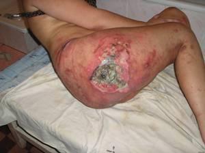
III stage- destruction of all layers of the skin. The subcutaneous fatty tissue also suffers, but the muscles are not damaged. A bedsore looks like a deep hollow wound.
IV stage- the most severe decubitus. Complete destruction of all layers of the skin, subcutaneous tissue, muscles, tendons. Bones and joints may be affected. People with fourth-degree pressure sores are at high risk of death from infectious complications.
Causes of bedsores

Healthy people do not experience bedsores because they are constantly on the move. Even during sound sleep, we unconsciously change our position in order to avoid prolonged squeezing of the same parts of the body. During the night, a person tosses and turns in bed up to 20 times.
Bedsores are formed in bedridden or sedentary patients with prolonged pressure on the soft tissues of the body. Due to pressure in the affected area of the skin, blood stops flowing, containing oxygen and nutrients necessary for tissue health. Without a constant blood supply, tissue becomes damaged and eventually dies. The poor blood supply also results in a shortage of white blood cells, the white blood cells that fight infection. After a bedsore has formed, bacteria infect it.
Possible causes of bedsores:
- hard surface pressure - bed or wheelchair;
- pressure from involuntary muscle movements - such as muscle spasms
- moisture, which can lead to a violation of the integrity of the upper layer of the skin (epidermis).
In addition, there are different types of mechanical effects that cause skin damage:
- surface pressure - pressing the skin against a solid surface by the weight of the body;
- shear and displacement of different layers of skin and soft tissues relative to each other occurs when a person slides down the bed or is lifted from the bed or wheelchair;
- friction, for example, of a mattress or clothing, against the surface of the skin.
The rate of formation of damage depends on the force of compression and the sensitivity of the skin. For example, in people with a predisposition, a bedsore that affects all layers of the skin can form in just one to two hours. However, in some cases, damage becomes noticeable only after a few days. There are various risk factors for pressure sores. They are described below.
Mobility restriction- any reasons that prevent moving the whole body or its individual parts. It can be:
- spinal injury;
- brain damage due to stroke or severe head injury;
- a disease that causes progressive damage to the nerves involved in body movement, such as Alzheimer's disease, multiple sclerosis, or Parkinson's disease;
- severe pain that makes it difficult to move the body or its individual parts;
- crack or fracture of a bone;
- recovery after surgery;
- coma;
- a disease that limits the mobility of joints and bones - for example, rheumatoid arthritis.
Improper nutrition- Healthy skin needs nutrients that can only be obtained from food. Reasons for a possible lack of nutrients in the diet:
- anorexia - a mental illness in which a person is obsessed with maintaining a low body weight;
- dehydration - lack of fluid in the body;
- dysphagia - difficulty swallowing.
Chronic illness, which disrupts blood circulation or increases the skin's predisposition to injury and damage. For example:
- diabetes mellitus type 1 and 2 - high blood sugar levels in this disease can disrupt blood circulation;
- peripheral vascular disease - restriction of blood flow in the legs due to the accumulation of fatty plaques in the vessels;
- heart failure - damage to the heart, in which it is not able to pump enough blood;
- renal failure - a violation of kidney function and the accumulation of dangerous toxins (poisons) in the body;
- chronic obstructive pulmonary disease (COPD) is a group of lung diseases that cause low levels of oxygen in the blood, which can make the skin more vulnerable.
Age over 70 years. There are a number of reasons why aging skin is more vulnerable to pressure sores, including:
- with age, the skin partially loses elasticity (the ability to stretch), which makes it easier to injure;
- decreased blood flow to the skin due to aging;
- with age, the layer of subcutaneous fat, as a rule, decreases, and the fat acts as a cushion - a shock absorber.
It is important to avoid putting pressure on areas that are prone to pressure ulcers and where they have already formed. Movement and regular position changes help prevent the development of bedsores and relieve pressure on existing ones. Lying patients in bed must be constantly moved. Usually this is done every 2 hours, on the recommendation of a doctor (if there is a high risk of pressure sores) - more often, up to once every 15 minutes.

Anti-decubitus mattresses and pillows
There are various special mattresses and pillows that help relieve pressure on vulnerable parts of the body. Anti-decubitus mattresses and pillows should be chosen together with your doctor. People with a predisposition to pressure ulcers and those who already have stage 1 or 2 bedsores should consider purchasing a custom-made mattress filled with foam to help relieve pressure on the body.
People with stage 3 or 4 pressure sores will need a more sophisticated mattress or system. For example, there are mattresses connected to a direct current of air, which automatically adjusts the pressure as needed.
Dressings and ointments for bedsores
Special dressings help protect bedsores and speed up healing. There are the following types of bandages:
- hydrocolloid - containing a special gel that stimulates the growth of new skin cells in the bedsore, while maintaining the surrounding healthy skin dry;
- alginate - made from algae and containing sodium and calcium, accelerating the healing process.
To speed up healing and prevent further tissue damage, special creams and ointments for bedsores can be used. A decontaminating cream is sometimes applied directly to the bedsore to kill bacteria. Antibiotic tablets are prescribed only for infected bedsores to prevent infection.
Treatment of bedsores - sanitation
In some cases, dead tissue may need to be removed to speed up the healing of a pressure sore. This is called sanitation - cleansing. If there is little dead tissue, the pressure ulcer is treated with special dressings and paste. Large areas of dead tissue must be removed mechanically. Mechanical methods for treating bedsores include:
- cleaning and irrigation under pressure - dead tissue is removed by jets of water under pressure;
- ultrasonic cavitation - sanitation of bedsores with the help of high-frequency sound waves;
- laser ablation - dead tissue is removed using high-energy light radiation;
- surgical debridement - cleaning the wound with surgical instruments.
Before treatment, the bedsore and the tissues around it are treated with a local anesthetic so that the debridement does not cause pain and discomfort.
Treatment with larvae
Alternative recovery method. The larvae are ideal for wound debridement, as they feed on dead and infected tissue without touching healthy tissue. They also help fight infection by releasing substances that kill bacteria and promote healing.
During the procedure, the larvae are attached to a bandage that is applied to the wound, and then this area is bandaged. After a few days, the bandage is removed, and the larvae are removed. The idea of maggot treatment seems abhorrent, but some studies have shown that this method of debridement may be more effective than traditional ones. However, this method of treating bedsores is not officially used in Russia.
Surgery to treat pressure sores
Third- or fourth-degree bedsores rarely heal on their own. In this case, an operation is required, which consists in cleaning the wound and closing it by suturing the edges (direct closure) or using tissue taken from a neighboring body area (skin flap plastic).
Surgery to close a pressure sore can be tricky, especially given that people with pressure sores are often already in poor health. The operation is associated with the risk of complications, such as the following:
- wound infection;
- tissue death of the sutured flap;
- bone infection (osteomyelitis);
- bleeding;
- deep vein thrombosis (blockage of a vessel by a blood clot).
Despite the risks, surgery is often necessary to avoid life-threatening complications such as blood poisoning and gangrene (rotting of living tissue).
Why are pressure sores dangerous?
Despite good care and treatment, stage III and IV bedsores can develop life-threatening complications. They are described below.
Purulent diseases of soft tissues, such as panniculitis - inflammation of the subcutaneous fatty tissue in the area of the bedsore and nearby tissues, necrotizing fasciitis - muscle fascia is involved in purulent inflammation, gas gangrene - destruction of soft tissues under the action of bacteria that live without oxygen. All these complications are very dangerous, they are manifested by an increase in body temperature, severe pain at the site of the lesion, swelling and redness. With purulent complications, urgent medical care is required: surgical treatment of the wound, a course of antibiotics. In severe cases, it may be necessary to amputate limbs.
Blood poisoning (sepsis)- the spread of infection in the blood and throughout the body. This is possible with severe bedsores in people with weak immunity. In the most severe cases, multiple organ infections can lead to a sharp drop in blood pressure (septic shock), a deadly complication. Blood poisoning is an emergency that requires immediate treatment in an intensive care setting, where bodily functions will be supported with medical devices until the infection can be cleared.
Joint and bone infection- septic arthritis and osteomyelitis. These complications can cause destruction of joints and bones. Antibiotics are used for treatment. However, in the most severe cases, surgical removal of damaged tissue may be required.
Prevention of bedsores
One of the most effective methods of preventing bedsores in bedridden patients is to regularly and frequently change the position of the body. If a bedsore has already appeared, regular movement will help relieve pressure on it and speed up the healing of the wound. Bedridden patients should change the position of the body at least once every 2 hours. Wheelchair-bound people should change position at least once every 15 to 30 minutes.
When a bedsore appears, it is important to try to reduce the pressure on it as much as possible so that the wound heals faster. If a person is not able to move himself, he should be assisted by a relative or a nurse.
For bedridden patients, anti-decubitus mattresses are used. Under those parts of the body that are most susceptible to compression, foam pillows of various thicknesses from 3 to 10 cm are placed. The bed should be filled with clean cotton linen. It is necessary to ensure that the sheet does not gather in folds, there are no crumbs and other objects in the bed that exert friction and pressure on the body. The underwear of a bedridden patient should be made of natural fabrics, without coarse seams and elastic bands.
It is necessary to strictly monitor skin hygiene, take daily water procedures with liquid soap. During washing, do not rub the skin. If necessary, use diapers or absorbent pads to keep the body dry and clean.
People who are prone to pressure ulcers should check their skin every day for signs of pressure ulcers, such as blemishes. Hard-to-reach parts of the body, such as the buttocks and soles of the feet, can be examined with a mirror. If you find any signs of damage, you should consult a doctor.
Nutrition for bedsores
A healthy, balanced diet that includes the right amount of protein and a variety of vitamins and minerals will help prevent skin lesions and speed up healing. If you have no appetite due to any disease, you should use the following tips:
- Eat small meals throughout the day instead of two or three large meals. You can make a schedule for eating, rather than waiting for the feeling of hunger. You need to consume enough nutrients.
- Before eating, you should not drink a lot of liquid, as this will create a false feeling of fullness.
- If swallowing is difficult, you can try special nutritional drinks or purees and soups.
- Vegetarians need to consume enough vegetable protein. Examples of protein-rich foods include cheese, yogurt, peanut butter, legumes, and nuts.
One of the most effective ways to prevent pressure ulcers for smokers is to stop smoking. Smoking leads to a decrease in oxygen in the blood, and also weakens the immune system, which increases the risk of pressure ulcers.
Which doctor should I contact for pressure sores?
If you or your relative has signs of pressure sores,. Your doctor will examine your skin and offer you treatment options. May require hospitalization. With the help of the NaPopravku service, you can call a surgeon at home.
Localization and translation prepared by site. NHS Choices provided the original content for free. It is available from www.nhs.uk. NHS Choices has not been reviewed, and takes no responsibility for, the localization or translation of its original content
Copyright notice: “Department of Health original content 2020”
All materials on the site have been checked by doctors. However, even the most reliable article does not allow taking into account all the features of the disease in a particular person. Therefore, the information posted on our website cannot replace a visit to the doctor, but only complements it. Articles are prepared for informational purposes and are advisory in nature.