Concave tibia. Injuries and structure of the tibia. First aid for fractures of the tibia
Anatomy
As we have already found out, a bone consists of two bones.
- Great tibia. It is located medially. This is a tubular long bone with two and three-sided body. The upper end, that is, the proximal epiphysis, together with the patella, forms the knee joint. The distal epiphysis is connected to the talus to form the ankle joint.
- Small tibia. It is located laterally. This is also a tubular long bone, however, it is much thinner than the tibia.
There is a large space between the first and second bone. In addition, the tibias are connected to each other by the tibiofibular joint in the region of the proximal ends.
If you make a slight increase, you can see that the intercellular substance of the bone tissue consists of thin plates. They differ from each other in thickness and shape, however, most of them exist in the form of hollow cylinders with different diameters. They are inserted one into one and form osteons. These plates are located depending on the direction of the blood vessels that run along the bone length. The transverse section shows that the osteons are concentrically arranged Haversian plates. The osteon has a cavity in the center called the haversian canal. Nerves and blood vessels pass through it.
The tibia is the strongest bone in the human skeleton. It is most affected when the body is in an upright position. It can withstand a load of up to 1650 kg, and this exceeds the usual load by 25 times. However, this bone is also distinguished by exceptional lightness, which is associated with its microscopic structure. It is covered with periosteum, which consists of an outer and inner layer. There are many vessels and nerves in the periosteum. It determines the innervation and nutrition of the bone.
With regard to the fibula, it is worth saying that it carries almost no tangible physical load. One of its main functions is that it is involved in the formation and development of the knee and ankle joints.
Damage
Fractures of the tibia are rare, but they can occur in both adults and children. Even without special medical knowledge, it becomes clear that damage to this part of the skeleton is quite dangerous. A fracture can affect one of the two tibia bones, or both bone elements can be damaged at once. A displaced fracture may occur. Any of these injuries requires quality first aid and effective treatment afterwards.
The bones of the lower extremities of each person experience great stress every day. This load becomes even greater in the following cases:
- Excess weight.
- Weak muscles.
- Impaired coordination of movements.
 If the bones do not cope with the functions assigned to them, they are destroyed. Fracture of the tibia is one of the most common types of fractures of the lower extremities. Such injuries occur for various reasons, therefore they differ in severity and nature. For example, direct damage results in fragments of one type, while indirect injury entails other types of fragments.
Most often, a fracture occurs due to a strong blow or fall. If a displacement occurs, this means that a strong blow was made to the knee joint in a bent form. The displacement of the condyles can occur both outwards and inwards. Oblique and helical injuries occur when, at the time of the fracture, a strong bend or rotation occurred in the lower leg, while the foot was in a fixed position. The situation is aggravated if there is a fracture of two bones at once. In this case, it is impossible to set the bone fragments.
Now we need to discuss two important points: how to determine a fracture of the tibia and what to do about it.
 If there is a fracture with a strong displacement, you need to determine its type. If this is an oblique plane, traction is used. Pins are inserted through the bone, after which an individual weight of the load is hung to stretch the leg. If a transverse fracture occurs, a metal plate must be applied. It is fixed with a plaster bandage. The fracture is then treated as a normal displacement. In the case of a fracture of both bones, much depends on the fragments that were formed as a result of it. If the fragments cannot be compared, a surgical operation is performed.
Prevention
All bones, including the tibia, must be protected. When cycling, skating or rollerblading, you need to use protective equipment, that is, shin guards, knee pads, and so on. All safety measures must be taken for the child. You must always follow the traffic rules.
In order for the tibia to be strong, it is necessary to deliver a sufficient amount of calcium to the body. It depends on nutrition. Caution and a healthy lifestyle can protect us from many injuries, so let's take care of the health of both our own and our children.
The tibia is part of the peripheral skeleton, which connects the bones of the thoracic and pelvic limbs. The tibia and fibula form the lower leg. Injuries to these parts of the skeleton immobilize a person for a long time and pose a threat to his health.
The structure of the tibia
As we have already found out, the tibia and fibula form the lower leg, and are located in its inner part. If we put our hand on the front of the leg (below the knee), then we immediately rest against the tibia. And on the outside of the lower leg is the fibula, which cannot be touched, since it is located in the thickness of the muscles. Consequently, these two bones are interconnected and form the ankle joint on one side, and the knee joint on the other. Thus, their structure determines the mobility and functionality of the lower extremities.
tibia
The tibia is located closer to the center in relation to the small bone. It is a tubular long bone, which is equipped with two epiphyses and a body. Her body consists of three edges that are triangular in shape:
- front;
- interosseous;
- medial.
These edges have three surfaces:
- back;
- medial;
- lateral.
The upper epiphysis, together with the patella, forms the knee joint. The lower part articulates with the talus and forms the ankle. The tibia is the most massive and stable bone in the human skeleton. She experiences the greatest stress when a person is standing, running or walking fast. In addition, this bone is very light because it has a microscopic structure, it is penetrated by multiple vessels and nerve endings.
Tibia
It is located on the outer (lateral) side of the lower leg. It is also a long tubular bone, but much smaller in shape and thickness. Consists of two epiphyses: upper and lower. The upper one goes into the knee joint, and the lower one goes into the ankle joint. As part of the ankle joint, it is called the lateral (outer ankle). Its main function is to stabilize the ankle joint. However, it practically does not carry any load, but is a place of muscle attachment.

Has three surfaces:
- back;
- medial;
- lateral.
These surfaces are separated by three ridges.
Injuries
Traumatization of the lower extremities occurs due to the large load on the joints that they experience every day when walking and moving. Injuries to the lower leg usually damage both bones.
In addition, in some cases, this load increases:
- overweight or obese;
- congenital anomalies of the skeletal system (in this case, the lower extremities);
- with a weak muscular apparatus;
- with impaired coordination of movements.

In these cases, the bones cannot cope with the load that is placed on them, which leads to injury. Such injuries occur for various reasons and, depending on this, differ in nature and severity. For example, with direct bone injury, fragments of one type are observed, and with indirect injury, another type.
Causes of damage to the tibia:
- swipe;
- car crashes;
- falling from a height;
- industrial injuries;
- excessive physical activity (for example, during professional sports).
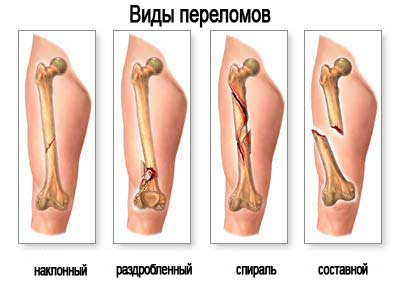
Fracture classification
Injuries to the lower leg usually damage both bones. Fractures of the body of the tibia are almost always accompanied by displacement of bone fragments. They are of the following types:
- Transverse. If only the tibia is fractured, then there is a stable damage to the bones without displacement of the fragments. If a small bone is damaged, then fragments are unstable.
- Helical. Observed when subjected to a torsional force, the damage is unstable and has a spiral shape.
- Oblique. As a rule, occur at an angle. Damage is unstable, with a tendency to increase displacement.
- splintered. They are characterized by strong instability and the formation of more than three bone fragments.
In addition to this classification, fractures are either closed or open. With closed fractures, there is no violation of the integrity of the skin.
When open, the skin is damaged, and broken fragments communicate with the external environment. This type of damage is also dangerous because the resulting wound can become infected.
Such injuries are far from rare, they happen both in adults and in children. You do not need to have special medical knowledge to understand that trauma to this anatomical segment is very dangerous and can lead to serious consequences.
Especially dangerous are injuries affecting damage to both bones. Indeed, in this case, a person is waiting for complete immobility and a long rehabilitation. Displaced fractures are possible, which also require a long recovery period.
Fracture symptoms
Symptoms are characterized by severe sharp pain, rapidly increasing swelling, bruising and bruising, and obvious shortening of the injured limb. The victim can not only walk, it is impossible to lean on and simply stand on the injured limb. As a rule, such fractures always occur with displacement of the fragments. The leg may take the wrong position and be turned in a certain direction: inward or outward (in relation to the knee). With an open fracture, there is damage to the skin through which bone fragments are visible.
The diagnosis is made with the help of radiographic examination, since one clinical picture is not enough. The study of the radiograph allows you to determine the number of fragments and the degree of their displacement, the presence of a fracture of both bones or only one of them, as well as the integrity of the knee and ankle joint. Also determine the integrity of blood vessels and nerves. For this, the victim is sent for a consultation to narrow specialists.
Treatment and first aid
The provision of first aid may affect the further treatment and rehabilitation of the victim. First of all, he is given analgesics and anti-shock therapy (in the presence of multiple injuries). Immobilization of the lower leg is carried out using a splint. Any object at hand (plywood, skis, boards) can act as a tire. When applying a splint, it is very important that its lower part covers the ankle joint, and the upper one ends at the top of the thigh.

With an open fracture, a tourniquet should be applied just above the wound to stop the bleeding. Be sure to treat the open wound with iodine, alcohol, brilliant green, or simply rinse with water if disinfectants are not at hand. All these actions are necessary to minimize infection of the wound.
Conservative treatment
Treatment in a medical institution can be both conservative and surgical. Treatment tactics depend on the degree and level of damage. For injuries that are stable and without displacement (which happens extremely rarely), a plaster cast is applied. For other types of damage, skeletal traction is used. The essence of this treatment is that a metal needle is passed through the heel bone, and a splint is placed on the leg.
Such treatment suggests two scenarios. Firstly, conservative treatment involves traction for 4 weeks, during which the bone fragments are fixed in the correct position. When a callus appears, the skeletal traction is removed and a plaster cast is applied for another two months. Secondly, after removing the bandage, the patient is prescribed rehabilitation: physiotherapy, massage and therapeutic exercises.
Surgical treatment
Surgical treatment is indicated for multi-comminuted fractures that are difficult to restore in the correct anatomical position with traditional conservative treatment. Surgical treatment involves the use of a variety of metal structures - plates, pins, rods. In addition, with such injuries, the use of the Ilizarov apparatus is indicated. The device allows you to restore the natural location of the fragments and their rapid fusion. It is used in the most difficult cases - with comminuted fractures with the formation of a bone defect. The period of bone fusion is approximately 4-6 months. The pace of recovery is individual and depends on the degree of damage and the complexity of the injury.
The lateral malleolus is the external bony stabilizer of the ankle joint.
The structure of the fibula
The body of the bone has a prismatic trihedral shape, curved backwards and twisted around the longitudinal axis. The fibula has three surfaces: posterior, lateral, and medial, which are separated from each other by three ridges.
The anterior edge has the shape of a sharp ridge and separates the lateral surface from the medial. The medial crest is located between the medial and posterior surfaces of the bone, and the posterior edge is located between the lateral and posterior surfaces. On the posterior surface there is a nutrient hole that extends into a distally directed nutrient channel. On the medial surface, the interosseous margin can be seen.
The upper epiphysis of the fibula forms the head, which, with the help of the articular surface, is connected to the tibia. The upper part of the head has a pointed shape and is called the top of the head. The head is separated from the body by the neck of the fibula.
The lower epiphysis of the bone forms the lateral malleolus. Its outer surface is well felt through the skin. On the medial surface of the lateral malleolus is the articular surface, with the help of which the bone is connected to the outer part of the talus. Slightly higher on the fibula is a rough surface that connects to the fibula notch of the tibia.
On the back surface of the outer ankle, you can see a tendon trace of the long peroneal muscle - the ankle groove.
Types of fractures of the tibia
Fractures occur at different levels of the fibula. Predominantly, the bone breaks in the region of the lateral malleolus. In turn, a fracture of the lateral malleolus of the lower leg occurs at its various levels. As a rule, a fracture of the fibula is accompanied by dislocation or subluxation of the foot, shortening of the bone, and rupture of the distal interosseous syndesmosis.
There are oblique, comminuted, transverse, spiral and fragmental fractures of the fibula.
The main symptoms of a fracture include:
Treatment of fractures of the tibia
The main goal of conservative treatment is the comparison and retention of bone fragments. A traumatologist performs a reposition, with the help of which the subluxation of the foot and the displacement of fragments are eliminated. If during the period of reduction of the fracture, the reposition was successful and the condition of the fragments is satisfactory, the foot and lower leg are fixed with a plaster cast or a special orthosis.
If the reposition does not give satisfactory results and the displacement of the fragments persists, surgical treatment of the fibula is prescribed, which consists of several stages:
- open reposition of bone fragments is carried out;
- subluxation of the foot is eliminated;
- bone fragments are fixed using implants (pin, screws, plate).
Anatomy of the fibula
The musculoskeletal system of the lower leg is represented by two bones - this is the tibia and fibula. They are the basis to which the muscles are attached and make the act of movement possible. Also important elements include cartilage, ligaments, blood vessels and nerves, which ensure adequate functioning of the limb. Due to the special structure of the lower leg, a person can both be stable in an upright position and perform active movements.
Anatomy allows you to study the structure of bones and soft tissue structures, the features of their connection, location, blood supply and innervation. It is worth noting that anatomy is considered a fundamental science, which is the basis for all other medical disciplines. Indeed, without knowledge of the normal structure of structures, it is impossible to identify their pathological changes.
What is bone made of
The fibula consists of a body (diaphysis) and two ends (epiphyses). It is located behind the tibia. She is much thinner than the latter, and her body is in the form of a three-sided prism. At the upper epiphysis is its head, which has a rounded shape. On its surface there is also a tubercle - this is the top of the head of the fibula. It is localized slightly behind and sideways on its rounded surface. Also, the tip of the head of the fibula has an articular part on its inner surface, which provides a connection with the external condyle of the tibia.
Below the head is its neck, which gradually passes into the main part - the body. It defines the inner, outer and back surface. Each of them is limited by the front, back and inner edge. Almost the entire length of the inner edge of the tibia to the outer edge of the fibula is the interosseous membrane. The rear surface has a feeding hole through which the feeding channel passes. In addition to its triangularity, this bone is somewhat twisted along the axis.
Towards the lower end, the fibula begins to thicken and form the outer malleolus. It can be easily felt through the skin. On the inner surface of the outer malleolus is the articular surface, which allows it to connect with the talus of the foot. Behind the place of their articulation is the fossa of the outer ankle, where the tendons of the lower leg are located.
The outer ankle is somewhat longer than the inner.
Leg muscles
The anatomy of the muscular apparatus of the lower leg has its own characteristics, since it is on the lower limbs that the greatest load falls. This causes a high degree of their development. The longest peroneal muscle and the short peroneal muscle have reached the greatest development in this area. All of the following muscles originate from the fibula.
The long extensor of the fingers from its place of attachment goes to the ankle, where it is divided into 4 tendons, which are attached to the middle phalanges of the II, III, IV and V fingers. The long extensor of the thumb departs in front in the middle third of the lower leg and, heading down, passes along the rear of the foot to the distal phalanx of the first finger.
The long peroneal muscle has several attachment points - this is the tip of the head of the fibula and the tibia. From them, the long peroneal muscle goes down, reaches the ankle and passes behind the outer ankle. Further along the calcaneus, it passes to the sole and is attached by the second end to the I, II metatarsal bones. Moving away from its place of attachment, the short peroneal muscle goes around the external condyle, and then goes along the outer surface of the calcaneus.
The short peroneal muscle is attached to the fifth metatarsal bone.
Ligament apparatus
The tibia and fibula are articulated with the help of the tibiofibular joint and syndesmosis. The articular surface of the first and the head of the second form the tibiofibular joint. Also, next to the tibiofibular joint, the articular capsule of the knee joint is fixed. It is strengthened in front and behind by the anterior and posterior ligament. It cannot be said that there is a lack of all kinds of movements in the tibiofibular joint, but nevertheless they are significantly limited.
The tibiofibular syndesmosis is a fixed joint, where the lack of movement is due to the peculiarities of the connection of the lateral malleolus with the tibial epiphysis. This connection is fixed by the anterior and posterior tibiofibular ligament. Both the anterior and posterior tibiofibular ligaments are located between the tibia and the lateral malleolus. Also, the lateral malleolus is the site of attachment of such ligaments as:
- anterior talofibular ligament;
- calcaneofibular ligament;
- posterior talofibular ligament.
Departing from their place of attachment, the anterior fibular and posterior talofibular ligaments pass to the foot and are fixed at the talus. The second place of their attachment is the calcaneus.
Innervation of the leg region
The anatomy of the nervous system in the lower leg area allows not only to study its structure. It makes it possible to understand the interaction of all elements. The innervation of the lower leg is provided by the following nerves.
It is the nervous system that unites individual anatomical structures, and its damage causes the absence of their functioning as a single organism.
Tibia: 3 parts
The tibia is located in the lower part of the leg. The tibia is an integral part of the skeleton of the lower leg. The tibia is a common name, in the skeleton of the lower leg are the tibia and tibia. Injuries to these bones significantly affect the deterioration of the musculoskeletal system and are very dangerous for health.
Tibia: fracture
The tibia is located inside the lower leg from the front side, this bone is the strongest of all human bones and can take pressure up to 1645 kg. The tibia is quite long, you can even roughly measure its length, from the knee to the ankle. The tip of the tibia is part of the knee joint, and with any body movements of a person, its work is involved, it is very important for the skeleton, since it is thanks to it that a person can take a vertical position, be stable and move around.
The components of the tibia are:
- The trihedral body of the bone itself;
- Upper epiphysis;
- lower epiphysis.
Injury to the tibia is common, even though the bone is very strong, and when it does, it can be very painful, whether it's slightly bruised or fractured. Fractures of the tibia are divided into three types: transverse, oblique and comminuted.
Such an injury should not be ignored, since not only is it unbearably painful, but there is a high risk of improper bone fusion and callus formation.
In case of improper fusion, in the future, an operation will be required, during such an operation, the doctor breaks the fused bone, removes calluses, attaches pins and applies plaster. The healing process is very long and painful, not to mention the rehabilitation process. In order to avoid, or at least reduce the risk of fracture, you need to know the following factors that put a person in a zone of predisposition to fracture of any bones of the lower extremities.
In case of a fracture of the tibia, you should consult a rheumatologist
- Overweight and obesity;
- Weakened, untrained muscles;
- Problems with motor coordination.
If in the first two cases a person can cope without the help of a doctor, but simply by bringing his body into a proper, healthy form, then the last point must be discussed with the attending physician. To protect children from any fractures and health problems, it is recommended to send them to sports sections or play sports together. In most cases, these types of fractures occur due to impacts or falls.
Fracture of the tibia and tibia
A fracture of the tibia most often occurs together with a fracture of the fibula, this "mechanism" practically does not break separately.
In most cases, such an injury occurs when:
- accidents;
- When a person falls from a great height onto a sufficiently hard surface;
- Engaging in active sports, such as skiing, mountain biking, sports riding on skateboards and snowboards, etc.
The cause can be any strong and sharp impact on the bone. The main thing is to correctly and in time determine that a fracture has occurred!
This injury is characterized by symptoms such as:
- Strong pain;
- Swelling of the limb, swelling of the fracture site and around it;
- Irregular shape of the lower leg, its curvature;
- The ability to move the lower leg itself, and not the knee joint.
There are two ways to treat a fracture of this kind: conservative, if there is no need to remove bone fragments and severe external damage to the tissues of the lower leg. In this variant, a fixator is placed for the patient to stretch and properly heal the bone, this lasts about 4 weeks, then they check whether everything has grown together correctly, using an X-ray, in a positive case, a plaster is applied and the patient walks with it for 2-3 months. Treatment can also be operative, it is used in cases of comminuted fractures, since it is simply not realistic to put all the fragments of the bone in place and put it correctly in a conservative way. This treatment option is characterized by the use of metal structures as auxiliary systems for restoring the patient's bone. As with conservative treatment, the patient is put in a cast.
Before choosing the type of treatment, in any case, X-rays are taken, and the larger the sides of the limb are illuminated, the clearer the injury and further treatment will be.
Long-term rehabilitation is necessary for high-quality restoration of the musculoskeletal system. The leg needs not only to be developed daily, but also to apply physiotherapy and exercise therapy, as prescribed by the doctor.
Tibia
This bone is also located in the lower leg, long and thin, has two “heads”, upper and lower, the latter is part of the ankle, it stabilizes the ankle joint. It connects to the tibia with an interosseous membrane. The structure is similar to the tibia, but there are important differences. The body of the fibula is slightly twisted and twisted initially, but it has a fairly simple structure. It is thin and not as strong as the tibia, but their "tandem" makes the lower leg resistant to external injuries.
If the tibia is damaged, an x-ray should be taken
The fibula has edges:
With the help of the thicker distal end, the bone forms the ankle.
Where is the tibia located
The fibula is located at the bottom of the human skeleton, or rather in the lower leg.
There are large and strong ligaments between the tibia and fibula. There is a hole on the back side of this bone, it exists in order for the vessels and nerves to enter it, they pass through the channel into the bone and interact with the rest of the channels of the human skeleton.
The main function of the fibula is the ability of the foot to rotate in different directions relative to the lower leg.
This is the most important function, but because of this feature, it is at high risk of being broken. The bone, although small and thin, should not be underestimated, it is very important for the skeleton, for its stability and ability to move.
fibula injury
The types of fracture of this bone completely coincide with the variants of a fracture of the tibia. Most often they break and are injured together. Since the force of injury passes in front and collides with the tibia, but after breaking it, the force is transferred to the fibula.
With an injury to the fibula, you can not self-medicate
- An open fracture is a fracture in which the bone extends beyond the muscular skeleton and skin, sticks out with a sharp edge and bleeds heavily, this fracture requires immediate surgical intervention and its treatment will take about six months. This is not only severe pain, but also a lot of stress for a person, it is not very pleasant to watch your leg in this form.
- A closed fracture is a more humane option for the patient's nervous system, but by its structure it is not always less dangerous. If there is no displacement and comminuted fracture, then the patient is lucky and the treatment will last not six months, but three months.
As after any fracture, the bone will never be the same and complete as it was before the injury, but with proper treatment and long and hard rehabilitation, it can restore its functions to almost full extent.
The first rule if a fracture is suspected is to turn to injuries. paragraph. There you need to make sure that they take an x-ray and clearly and clearly explain the type of fracture, the treatment technique and the recovery period. There is no need to be afraid to ask questions to doctors for fear of seeming stupid, a person, especially traumatized and prone to a stressful situation, more than ever needs support and understanding. Having received such an injury, you need to prepare yourself for a long recovery, special exercises and treatment, be patient and desire to recover as soon as possible.
Where is the tibia (video)
Such an event as a fracture is always unpleasant and at the wrong time. But if it happened, for one reason or another, you need to pull yourself together, endure the pain (doctors prescribe painkillers) and tune in to recovery. How much to walk in a cast and therefore fragmentation occurred, the doctor will explain.
The fibula: where is it located, functions, fracture options and their treatment
The fibula is represented by an elongated tubular formation. The bone is represented by a body, or diaphysis, and two peaks, called epiphyses. The lower fragment, called the lateral malleolus, is involved in the creation of the ankle joint. The lateral malleolus acts as a kind of stabilizing factor in the joint located between the lower leg and the foot.
Anatomy and position relative to other bones
The musculoskeletal system (ODA) in adults is represented by active and passive parts. The active component includes muscles, ligamentous apparatus. The passive fragment is indicated by a skeleton consisting of bones and their joints. In the body of an adult, this part is represented by 208 bones. In order to properly redistribute the mass of the human body in the process of life, the inner part of the bones is hollow. With the help of this, the weight of the skeleton is less in comparison with the total mass, however, despite this, the structure of the bones is strong, which allows the body to function adequately to the loads applied.
To appreciate the physiological significance of the tibia, it is necessary to understand their topography. The fibula is located in the lower part of the skeleton (region of the legs), between the thigh and foot, in contact with the tibia. From above, the tibia is limited by the knee joint, from below by the ankle joint. The small bone connects to the foot through the lateral malleolus through the ankle joint. Large ligaments are located between the tibia.
In accordance with the length, 3 parts are distinguished in the fibula: the diaphysis (body) and 2 epiphyses (upper, lower fragment). The body of the bone is bent posteriorly and twisted along the axial direction. The diaphysis is represented by a prism and consists of three faces: medial, lateral and posterior. Each of the faces is separated by a ridge. The medial and lateral edges are separated by an anterior protrusion, the internal (medial protrusion) subdivides the medial and posterior sides of the bone, and the posterior crest is located between the posterior and lateral sides.
On the back of the MBC there is an opening for the exit of blood vessels and nerves. From this hole, a special channel extends distally into the bone, communicating with the channels of other areas of the skeleton through holes. On the inner side between the bones is a delimiting edge. The upper epiphysis, represented by the head, is in contact with the tibia on its articular side. The top is pointed. The head is connected to the diaphysis of the fibula through the neck.
One of the most important formations of the fibula is the feature of topography and interaction with the bones of the foot and lower leg through the lower epiphysis. The distal part of the bone is often referred to as the lateral malleolus. This ankle is easily palpable through the skin when the foot is flexed forward.
On the inner side of the lower epiphysis is the articular side, which connects the talus and the lateral malleolus. Slightly higher in the fibula there is a slight roughness, connecting with the fibular notch in the tibia. Posteriorly on the fibula there is an ankle groove. The tendon of the peroneal muscle passes through this depression.
For the prevention and treatment of diseases of the JOINTS and SPINE, our readers use a new NON-SURGICAL treatment based on natural extracts, which..
Impact on functions in the musculoskeletal system
The leading function that the fibula performs, laid down in the process of ontogenesis, is the provision of rotation in the ankle. Rotation in this case is a turn to the right or left of the lower leg and foot in relation to each other. Given the anatomical structure, location, under the influence of a strong traumatic aspect, the bone tissue is prone to fractures.
Joint problems - a direct path to disability!
Stop enduring this joint pain! Write down a proven prescription from an experienced doctor.
Usually, the fracture first appears in the tibia, as it takes on the leading stress when walking. Massive injuries or strong local effects of a negative factor can also cause damage to the tibia, often with rupture of soft tissues, displacement of bone fragments. Fractures occur in various parts of the fibula. Most often observed in the lower epiphysis.
Options for fractures of the tibia:
- transverse;
- oblique;
- spiral;
- splintered;
- fragmentary.
Fractures are usually combined with subluxation and dislocation of the foot, tearing of the distal syndesmosis between the tibia, shortening of the bone. To understand that a fracture of the entire or fragment of the fibula has occurred, it is necessary to note a number of characteristic symptoms, the main of which are pain at the site of the lesion, which increases with palpation and making movements in the ankle or applying a vertical load, swelling.
The pain is noted constantly and increases when walking or standing. These symptoms usually occur after a leg injury or fracture. To restore bone function to the full, it is necessary to consult a traumatologist as soon as possible.
Briefly about therapeutic measures and healing time
Treatment of fractures of the tibia is carried out conservatively or surgically. First, proceed to non-operative intervention. The conservative technique is based on the comparison of disconnected fragments of bone tissue and their subsequent retention. The primary moment in the tactics of treatment, the traumatologist should carry out the reposition of the fragments, thereby excluding further dislocation of the MCD and subluxation or dislocation of the foot. Upon successful completion of the reposition, confirmed by the results of an X-ray examination, the ankle is closed with a plaster mass or an orthosis.
In a situation where the docking and fixation of bone pieces did not give the necessary results, a surgical intervention is prescribed, represented by a number of stages:
- comparison of bone tissue fragments in an open way: incision of soft tissues, moving muscles together with vessels and ligaments, creating access directly to the fracture point;
- elimination of subluxation, dislocation of the foot;
- fixation of bone fragments using implants: pin, screws or plate;
- closure of the fracture site with a plaster mass to immobilize the ankle and create the best conditions for bone restoration.
After the surgical intervention, the patient must undergo a period of rehabilitation. The terms of fusion of the fibula are individual, and in uncomplicated cases correspond to 2-3 months. When multiple bone fractures were noted, and there was also a burden in the anamnesis (somatic pathology in the stage of compensation and decompensation), the fracture in the fibula continues to heal for six months. In order to accelerate the overgrowth of the fracture, to recreate the functions, the patient is prescribed therapeutic exercises and massage. Not in the acute period, treatment is supplemented with physiotherapeutic intervention.
Most people who are faced with fractures of the bones of the lower extremities, especially the tibia, which plays an important role in the development of the ankle joint, are concerned about the further consequences and forecasts of qualified specialists.
The result of treatment depends not only on the correct comparison and fixation of fragments. It is extremely important that the patient strictly follow all the recommendations of the doctor. It is especially necessary to protect the fracture area from excessive physical activity during the rehabilitation period and after. The sooner the patient seeks qualified help from the moment of leg injury, the greater the likelihood of successful treatment and complete rehabilitation.
Sometimes after a bone fracture, conservative or surgical interventions, the following consequences may occur:
- the appearance of dysfunctions in the ankle joint;
- tissue swelling and pain at the site of a regular lesion;
- deforming arthrosis and osteochondrosis;
- weather sensitivity.
So that problems in movement do not arise after a bone or ankle fracture has occurred, it is necessary to take care of the legs. If the injury still occurs, it is necessary to urgently seek an appointment with a traumatologist.
After a fracture, the site of the lesion should be protected throughout life and not subjected to greater physical exertion in the future.
Get the book "17 Recipes for Delicious and Inexpensive Meals for Spine and Joint Health" for free and start recovering effortlessly!
Where is the fibula located? In what cases does it fracture?
The human tibia is the smaller bone of the lower leg; when walking, it practically does not carry any tangible physical load. One of the main functions of this part of the lower limb is participation in the development and formation of the ankle and knee joints. Even though the bones of the legs are more massive than the skeleton of the hands, their injury is quite common among the population. Fracture of the fibula in most cases occurs simultaneously with the tibia, which is fraught with complications such as displacement of bone fragments and osteomyelitis. However, if only the fibula breaks, the treatment will be faster and more effective.
Symptoms of a femur fracture:
- strongly pronounced bone displacement;
- sharp pain when walking;
- swelling or hematoma in the area of injury;
- visual discrepancy between the length of the leg when the fragments are displaced.
Risk factors for a tibia fracture:
- lack of vitamin D and calcium, especially in the elderly;
- fragility of bones, mainly in young children;
- excessive load on the lower limbs, in particular in athletes;
- acquired diseases that provoke bone fragility;
- a strong blow, such as in a car accident.
In people involved in sports, fatigue fractures are common, which are very rarely open, so the fibula recovers quickly enough. In this case, the fracture is a small crack that develops over time. At the same time, the tibia swells and hurts a lot, and subsequent healing occurs without surgical intervention. To restore normal leg function, a cast is applied for a period of one and a half to two months.
Often, the fibula breaks in babies from one to three years old as a result of a fall from a height, as a rule, open fractures do not occur. The child develops swelling and severe pain, he begins to react painfully to any touch and refuses to rise to his feet. Since it is not always possible to take an x-ray, a bone scan becomes an ideal option for examining the bone. When a fracture is confirmed, a short bandage with plaster is applied to the child for an indefinite period, which depends on the personal characteristics of the recovery. As a rule, a complete cure occurs faster than in an adult, which is explained by an accelerated metabolism.
In severe cases of fracture of the fibula, both with closed and open injuries, surgery is necessary, followed by fixation of the ankle. In this case, a frame apparatus or fixing the bones with special pins can be assigned. If an infection has joined the fracture during injury, it is even possible to amputate a certain part of the leg. In order for the fibula to recover faster, it is advisable to prescribe therapeutic and prophylactic gymnastics. It must be remembered that a fracture is a temporary injury, which in most cases can be effectively treated, after which a person can walk as before. Therefore, do not despair and try to protect your legs from repeated damage.
Tibia
2-lateral condyle of the tibia;
5-tuberosity of the tibia;
10-articular surface of the ankle;
12-lateral malleolus (machotibia);
13- articular surface of the ankle (lateral);
14-body of the fibula;
15-medial (interosseous) edge;
2-upper articular surface;
4-posterior intercondylar field;
6-apex of the head of the peroneal bone;
7-head of the fibula;
8-body of the fibula;
9-medial (interosseous) edge;
10-articular surface of the ankle (fibula);
11-fossa of the lateral malleolus;
12-groove of the lateral malleolus;
13-articular surface of the medial malleolus;
15-ankle sulcus (groove of the medial malleolus);
16-medial edge of the tibia;
17-body of the tibia;
18-lateral (interosseous) edge of the tibia;
19-line soleus muscle.
The fibula is a long thin tubular bone. It consists of a body and two ends, respectively, upper and lower. The body of the fibula has a trihedral prismatic shape, twisted around the longitudinal axis and curved backwards.
Atlas of human anatomy. Akademik.ru. 2011 .
See what "Fibula" is in other dictionaries:
FIBIBLE BONE - A long, thin, externally exposed bone of the lower limb of four and bipedal vertebrates, including humans. It articulates with the TIBIUS just below the knee; its lower end forms a ledge on ... ... Scientific and Technical Encyclopedic Dictionary
Fibula - The fibula is a long, thin, tubular bone. It consists of a body and two ends, respectively, upper and lower. The body of the fibula has a trihedral prismatic shape, twisted around the longitudinal axis and curved backwards. Three surfaces ... ... Wikipedia
tibia - tibia / tibia (large and small) One of the two parallel bones of the lower leg ... Dictionary of many expressions
TIBIUS BONE - TIBIUS BONE, the inner, larger of the two lower bones of the leg. At the knee, it connects to the THIGH, or upper leg bone, below it passes into the ankle. Its lower end forms the ankle bone protruding from the inside of the leg. see SMALL ... ... Scientific and Technical Encyclopedic Dictionary
BONE - BONE. Contents: I. HISTOLOGY AND EMBRYOLOGY. 130 II. bone pathology. sh III. Clinic of bone diseases. 153 IV. Bone operations. Yub I. Histology and Embryology. The structure of K. of higher vertebrates includes ... ... Big Medical Encyclopedia
The fibula is (fibula, fibula) one of the two bones of the lower leg (see) ... Encyclopedic Dictionary F.A. Brockhaus and I.A. Efron
MUSCLES - MUSCLES. I. Histology. In general morphologically, the tissue of the contractile substance is characterized by the presence of specific differentiation in the protoplasm of its elements. fibrillar structure; the latter are spatially oriented in the direction of their contraction and ... ... Big Medical Encyclopedia
Birds - "Bird" redirects here; see also other meanings. Birds 18 ... Wikipedia
FOOT - foot (pes), the distal part of the hind limb of terrestrial vertebrates, articulated at the top with the lower leg and acting as a supporting element. S. consists of 3 departments: tarsus, metatarsus, and phalanges of stingers. In most animals, reliance is made on ... ... Biological Encyclopedic Dictionary
Skeleton - (from the Greek. skeletos, literally dried up) a set of hard tissues in the body of animals and humans, giving the body support and protecting it from mechanical damage. There are external and internal S. In most invertebrates S. ... ... Great Soviet Encyclopedia
Fibula
The fibula (fibula) is a long and rather thin tubular bone of the lower leg. It is much thinner than the tibia. This bone consists of a body and two - upper and lower epiphyses. The primary function of the fibula is to rotate the tibia and foot, allowing the foot to rotate left and right. Such movements are due to the rotation of the lower leg, i.e. rotation relative to each other of the fibula and tibia. The body of the fibula, located between these two epiphyses, has a trihedral prismatic shape, it is bent backwards and twisted around the longitudinal axis. The fibula has lateral, medial and posterior surfaces, which are separated from each other by edges (ridges). The lateral surface is separated from the medial by the anterior edge, which is the sharpest ridge. The medial crest separates the posterior and medial surfaces. The posterior edge separates the posterior and lateral surfaces of the fibula. The posterior surface of the bone has a so-called nutrient foramen, which extends into a distally directed nutrient canal. The medial surface of the fibula contains the interosseous margin.
The head of the fibula is formed by its superior epiphysis. On the head of the bone there is an articular surface, which is necessary for connection with the tibia. Also on the head there is its tip, which has a pointed shape, separated from the body by the neck of the fibula. The lower epiphysis makes up the lateral malleolus, the outer surface of which is perfectly palpable through the skin. The articular surface of the ankle is located on its medial surface. The articular surface of the ankle connects the fibula to the outer surface of the talus. The rough surface, which is located above, connects the fibula to the fibula notch of the tibia. The posterior surface of the lateral malleolus has an ankle groove, which is quite shallow and is a trace of the tendon belonging to the long peroneal muscle.
The fibula is connected to the tibia by an interosseous membrane.
blood supply
The blood supply to the fibula in the middle third is carried out by a large nutrient vessel from the peroneal artery. Also, the supply comes from the periostenium, which receives many small vessels from the peroneal artery. The proximal head and epiphysis are supplied by a branch of the anterior tibial artery.
The structure of the human fibula
The lower leg, that is, part of the lower limb of a person, consists of the following bones: the tibia and the fibula. Muscles are attached to these components of the human body. The fibula itself consists of a long, thin, somewhat twisted body and two expanded ends. The upper end is called the head of the fibula, and due to its peculiar articular surface, it is attached to the tibia. This connection is made by an interosseous membrane. And the lower end is an ankle that enters the ankle joint. Such is the anatomy of this part of the human lower leg.
It is thanks to the fibula that the lower leg, as well as the human foot, can rotate. But this process occurs as a result of the rotation of the two bones of the lower leg relative to each other. We owe our mobility to these bones. According to the anatomical atlas, the fibula is located in the same place where the tibia is located, that is, in the lower leg.
What damage is the fibula prone to?
There are several types of damage to this bone.
When the fibula is fractured, the integrity of the body itself of this element of the human body is violated. It is located in the lower leg and usually breaks along with the tibia. The causes of fractures can be: traffic accidents, various domestic injuries, falls, bumps. People involved in extreme sports are more likely than others to break the fibula. Even this part of the lower leg is sometimes subject to fractures due to the lack of a balanced, full of vitamins and calcium, nutrition in the elderly.
Dr. Bubnovsky: “A penny product No. 1 for restoring normal blood supply to the joints. Helps with the treatment of bruises and injuries. The back and joints will be like at 18 years old, it is enough to smear once a day. »
The main types of fractures of the tibia.
- Fracture with displacement of bone particles.
- Fracture without any displacement.
- With the presence of fragments or without them.
- According to the very nature of the fracture: oblique or transverse, fragmentary or spiral.
- Depending on the blow that hit the bone: direct or indirect.
Types of symptoms in a fracture of the fibula.
- Sharp pain at the site of the injury.
- Swelling on the surface of the lower leg or even the foot.
- Obvious signs of hematoma.
- Somewhat deformed appearance of the limb itself.
- Muscles are pulled up to the injury and create the effect of shortening the leg.
- Leg numbness.
- Difficulty walking.
First aid for fractures of the tibia
When a bone is broken, you need to give a person a painkiller and be sure to immobilize the leg. It is impossible to treat a bone fracture on your own without medical qualifications. The victim must be sent to the clinic for an appointment with a doctor. To do this, you need to call an ambulance or take a taxi to the hospital.
Who diagnoses a fibula fracture?
A specialist in the treatment of fractures of the fibula is a traumatologist. The doctor first interviews the patient about how the injury was received. Then the doctor will require you to pass all the necessary tests and take an x-ray of the lower leg. Only after a detailed study of the nature of the injuries, the doctor will begin to treat the patient. After all, the fibula is treated, based on its anatomy, only by a traumatologist.
How is a hip fracture treated?
The doctor is involved in helping the patient, depending on the nature of the fracture. When the bone sticks out, sticks out, hurts a lot, then these are symptoms of a serious fracture, for the treatment of which an operation is needed. If no displacements are found on the x-ray, then the patient is simply put on the leg with a cast.
If parts of the bone come off, surgery will be required. With the help of special needles, the doctor will return the bones to the correct position. And metal structures will help fix the bone.
Also, a surgical operation is resorted to if the patient has an open fracture of the fibula, or with a significant crushing of this part of the lower leg. The doctor first restores the very shape of the bone, applying broken particles to each other. Then he fastens the parts of the bone together with special screws or plates.
How long does a fracture take to heal?
There is no one period of time during which all fractures of the fibula would be restored. Depending on the nature of the injury, as well as on the severity, on the age of the patient, on the qualifications of the attending physician, various injuries heal in different ways.
It can be argued that within two or three months there will be a fusion of the bones. The very same callus appears after six weeks. More severe injuries are restored after six months.
How is rehabilitation after fractures?
Four months after the complete healing of the broken bone, the rehabilitation process should begin. In time, it can drag on for six months or even more. It all depends on the severity of the fracture.
Types of rehabilitation for fractures of the tibia.
- Performing a scientifically developed complex of therapeutic exercises, which will help to “develop” a sore leg and bring it to mobility.
- Massages performed by professional manual therapists.
- Water procedures in swimming pools.
- Homemade baths from medicinal injuries.
- Independent rubbing of therapeutic ointments and creams.
- Gradual increase in the load on the sore leg under the supervision of the attending physician.
- Stretching of the knee joint.
In the event that the patient turned to the attending physician in time, and professional assistance was provided to him, then it will be easy to restore the working capacity of the leg. And after completing the rehabilitation course, the patient will be able to return to their usual and normal life in just six months.
- The bone may not heal properly.
- The wound may become infected.
- Nerves or blood vessels in the lower leg may be damaged.
- Thrombi are formed.
- The leg is bent.
All these unpleasant moments should be corrected. And only an experienced doctor can deal with problems. In some cases, he will prescribe a second operation.
For simple fractures and small cracks without displacement of bone particles, doctors do not use radical, but more conservative treatment. It consists in immobilizing the leg with a cast or splint. A splint is applied if there is significant swelling that does not allow a plaster cast to be applied to the swollen leg. Instead of a tire, a splint is sometimes used. But as soon as the edema decreases, the patient is immediately put in a cast.
Of course, the doctor performs all these procedures only after receiving an x-ray, indicating the nature of the damage to the leg. A patient with simple injuries of the fibula should be in a cast for about three weeks. After that, he is sent again by the attending physician for an x-ray. Based on the results of the wound healing process obtained with the help of a picture, the doctor additionally prescribes one or another treatment for his patient.
Consequences of fractures and prevention
Whatever the fracture of the fibula, it will almost always have consequences. Albeit not very complex, sometimes insignificant. But you should always pay attention to them. And in case of detection, seek help from a qualified doctor. After all, a slight pain in the lower leg can be a signal for a more serious disease. If you ignore it, then soon all sorts of disorders can occur in the human body, which will soon lead to serious illnesses.
And as a preventive measure, it is necessary to choose the right comfortable shoes for walking. Try not to walk in high heels. Protective equipment must be worn when playing sports. Do not expose your body to heavy physical exertion, which would lead to damage to the bones of the lower leg. Avoid in old age sports such as figure skating, skiing, roller skating. In winter, during ice, try to use shoes with non-slip soles. Carefully behave in transport, follow the rules of the road.
Other diseases of the fibula
But the fibula can be subject to various diseases. The most common is periostitis. It occurs as a result of advanced varicose veins. At the initial stage, the skin of the leg is not affected by any changes. But when palpated, the patient complains of unpleasant painful sensations.
The doctor starts treating the patient with periostitis based on the indications of the x-ray, tests and ultrasound scanning. The patient is prescribed medications, and he should also massage the sore leg, do rubbing. At home, this disease is not recommended to be treated. The patient needs specialist care. For a while, it is better to immobilize the leg.
Another disease of the fibula is osteoporosis. If you carefully consider the structure of this bone, you can find that it consists of a compact and spongy tissue. As a result of osteoporosis, the compact and spongy substance is destroyed. The bone becomes more hollow, and therefore brittle. Symptoms of the disease: pain in the lower leg, discomfort when walking. This disease is treated with the help of medications that are rich in calcium and phosphorus. And as a prevention of this disease, you should eat as much milk, cheese, fish as possible.
Osteomyelitis of the fibula is also a serious disease. This is a severe purulent, as well as infectious inflammation. Osteomyelitis affects all elements of the tibia. The cause of this disease is the penetration of dangerous microorganisms.
The disease develops against the background of immunodeficiency, as well as diabetes mellitus or a fracture of the fibula. This disease affects not only children, but also adults. The patient's body temperature rises sharply, the skin in the shin and knee area turns red, the person suffers from unbearable pain.
Treatment of osteomyelitis is carried out only in the hospital by a professional doctor: a surgeon or a traumatologist. Diagnose this disease with the help of x-rays, tests and computed tomography. You can’t open abscesses at home, because this can lead to sepsis and serious complications. In the hospital, the patient is examined by a surgeon. During the operation, the purulent focus is opened and eliminated. Medical preparations achieve a complete recovery of the patient.
The fibula is also prone to osteosarcoma. And this disease belongs to the category of the most dangerous diseases. As a result of its development, a malignant tumor is formed in the bone. At the initial stage, the disease almost does not manifest itself. A person attributes slight pains in the lower leg to the account of rheumatism. But he is wrong. The problem is much more serious. And after a few weeks, swelling appears, the pain becomes unbearable, metastases develop. Treatment for osteosarcoma involves surgery to remove the tumor. After that, the patient is prescribed a course of chemotherapy.
The diagnosis of this disease is carried out in the clinic, prescribing tests, x-rays, bone scans to the patient. A biopsy of tissue taken from the affected area of the fibula is performed. Previously, limbs affected by this disease were amputated. And the patients themselves did not live even five years after the operation. But now doctors have modern drugs in their arsenal. Thanks to new medicines, the percentage of patients who, even after the removal of metastases, continue to live for more than five years, has significantly increased.
Osteosarcoma affects young boys and girls. Most often it occurs at age. After fifty years, this disease is rare. Osteosarcoma can be caused, for example, by chemotherapy as a result of another cancer. Also, the disease can be activated after a bone fracture. The impetus for its development is osteomyelitis or Paget's disease.
Diseases that affect the fibula greatly weaken it. Sometimes the causes of fractures can be minor physical exertion, as a result of which the fibula breaks.
As a prevention of diseases of the fibula, it is recommended to eat a lot of fiber and calcium. Green vegetables help inhibit the development of pathogenic bacteria. Meat, milk, fish, cheese - these foodstuffs should always be on the table of a person. But in order not to get sick with dangerous ailments, it is necessary to lead a correct lifestyle.
A healthy lifestyle involves giving up smoking, alcoholic beverages, and drugs. The processes occurring in the human body can fail precisely because of the use of toxic substances. Everything in the human body is interconnected. And an ordinary cigarette can subsequently cause a sudden complication in the body, leading to the development of a malignant formation in the fibula.
How to forget about pain in the joints ...
Joint pain limits your movement and life...
- You are worried about discomfort, crunching and systematic pain ...
- Perhaps you have tried a bunch of folk methods and medicines, creams and ointments ...
- But judging by the fact that you are reading these lines, they did not help you much ...
The fibula is a tubular, thin and long bone of the lower leg. It consists of a body and two epiphyses, respectively, the upper and lower. The distal or lower end of the bone is an important component of the ankle joint and is called the lateral or lateral malleolus. The lateral malleolus is the external bony stabilizer of the ankle joint.
The structure of the fibula
The body of the bone has a prismatic trihedral shape, curved backwards and twisted around the longitudinal axis. The fibula has three surfaces: posterior, lateral, and medial, which are separated from each other by three ridges.
The anterior edge has the shape of a sharp ridge and separates the lateral surface from the medial. The medial crest is located between the medial and posterior surfaces of the bone, and the posterior edge is located between the lateral and posterior surfaces. On the posterior surface there is a nutrient hole that extends into a distally directed nutrient channel. On the medial surface, the interosseous margin can be seen.
The upper epiphysis of the fibula forms the head, which, with the help of the articular surface, is connected to the tibia. The upper part of the head has a pointed shape and is called the top of the head. The head is separated from the body by the neck of the fibula.
The lower epiphysis of the bone forms the lateral malleolus. Its outer surface is well felt through the skin. On the medial surface of the lateral malleolus is the articular surface, with the help of which the bone is connected to the outer part of the talus. Slightly higher on the fibula is a rough surface that connects to the fibula notch of the tibia.
On the back surface of the outer ankle, you can see a tendon trace of the long peroneal muscle - the ankle groove.
Types of fractures of the tibia
Fractures occur at different levels of the fibula. Predominantly, the bone breaks in the region of the lateral malleolus. In turn, a fracture of the lateral malleolus of the lower leg occurs at its various levels. As a rule, a fracture of the fibula is accompanied by dislocation or subluxation of the foot, shortening of the bone, and rupture of the distal interosseous syndesmosis.
There are oblique, comminuted, transverse, spiral and fragmental fractures of the fibula.
The main symptoms of a fracture include:
- pain when probing the outer ankle;
- edema;
- pain with axial load on the bone;
- pain in the ankle joint when moving.
Treatment of fractures of the tibia
The main goal of conservative treatment is the comparison and retention of bone fragments. A traumatologist performs a reposition, with the help of which the subluxation of the foot and the displacement of fragments are eliminated. If during the period of reduction of the fracture, the reposition was successful and the condition of the fragments is satisfactory, the foot and lower leg are fixed with a plaster cast or a special orthosis.
If the reposition does not give satisfactory results and the displacement of the fragments persists, surgical treatment of the fibula is prescribed, which consists of several stages:
- open reposition of bone fragments is carried out;
- subluxation of the foot is eliminated;
- bone fragments are fixed using implants (pin, screws, plate).
The tibia is one of the key bones in the human body. It is extremely large and is located medially compared to the lower leg bone. In its upper part, it connects to the femur. Thus, the knee joint is eventually formed. In the lower part it adjoins the talus.
Body of the tibia
The tibia is a regular trihedron with three distinct edges.
The first edge is the front. It can be felt through the skin and, if desired, palpated. In its upper part there is a characteristic tuberosity. It is in this place that the quadriceps femoris muscle joins the tibialis.
The second edge is interosseous. It is somewhat deployed in the direction of the fibula. At the same time, it is very sharp.
The last, third, medial edge is also called the middle edge by physicians. It has a characteristic rounded shape.

The structure of the tibia
At its end, the tibia forms a pair of thickenings that serve to attach muscles. In anatomy, they are called condyles. There are two of them in the tibia - lateral and medial.
On the side that is closer to the human thigh, the condyles are equipped with slightly concave platforms, which are necessary for connection with the same condyles of the femur. The articular surfaces of the condyles are separated by a kind of elevation, which has two tubercles. They are needed for reliable fastening of the articular ligaments.

Trauma to the posterior edge of the tibia
The posterior edge of the tibia is extremely susceptible to fractures and injuries. Injuries of this kind occur in a third of all victims of ankle fracture.
Most often, according to the experience of traumatologists, a bone fragment is located in the back, possibly on the lateral side of the surface. The connection of bone fragments after a fracture for their better fusion in the case of fractures of the tibia is carried out only when the size of the fragment does not exceed a quarter of the entire surface of the joint.
Doctors distinguish several types of damage to the posterior lateral edge of the tibia. Firstly, in the presence of a fragment of sufficient size, one speaks of a contour fracture of the posterior ligament.
Second, the condition of the fragments within the joint itself.
Thirdly, if the surface of the tibia itself is depressed during the fracture, then most often it is not possible to make an accurate diagnosis using only one initial x-ray. These are the most complex cases that require detailed and scrupulous study. Often, as a result of indentation, obstacles are formed, due to which it is not possible to quickly and effectively set the bone.

Right tibia
For clarity, consider what the tibia is. The right one, upon detailed examination, consists of 9 components.
At the heart of the intercondylar eminence. To the right and slightly below it is the medial condyle, and even lower and in the center is the tuberosity of the tibia.
An important component of the structure is the interosseous edge, under which the lateral surface is located, as well as the anterior edge, under which the medial surface is located.
The tibia ends with the medial malleolus. Anatomy today has carefully studied all the details of the structure of the human body, which today greatly simplifies the process of diagnosis and treatment.
Another component that cannot be ignored is the lateral condyle. It is located in the upper left side of the tibia.

Tibia by department
Considering the sections of the tibia, the main attention should be paid to the proximal section. It includes the upper part of the bone, which is directly involved in the formation of the knee joint. This department consists of two condyles. One is external, the second is internal, as well as the metaphysis. If during the damage the fracture line affected the articular surface of the tibia, then traumatologists call it articular.
Fractures of this part of the tibia can be mild (they are also called low-energy), for example, obtained by falling from a small height. As well as more complex or high-energy ones, for example, with a strong mechanical blow to the knee area - during a football match or a collision with a car.
The second cases are more dangerous, because as a result of such injuries, the likelihood of a large number of bone fragments is high. In any case, a fracture of this bone clearly belongs to the category of severe injuries and requires professional and qualified treatment. Most often, you can not do without surgery to splice broken bones and shift the resulting fragments. In this case, the tibia is fastened with screws, sometimes plates are added to them.
If it is precisely established that the fracture is intra-articular, then it is very important in this case to thoroughly restore the damaged surface, eliminating the displacement of bone fragments.
Otherwise, it is fraught with complications, the most common is post-traumatic osteoarthritis of the knee joints.

Intercondylar eminence
Another severe injury occurs when the intercondylar eminence of the tibia suffers. Such injuries are extremely painful and unpleasant. Most often they occur under the influence of indirect mechanical trauma. For example, when the impact is from behind or in front of the proximal leg, which is in a bent position. As a result, the cruciate ligaments are stretched to the limit, and the bone is torn off. Another cause of such fractures is excessive abduction or hyperextension.
The first signs of just such damage are sharp pain and swelling in the area of the knee joint. As a rule, the injury is the cause. Often such injuries occur in athletes from contact sports. The patient cannot fully extend the leg, most have a drawer symptom. True, it is not always possible to install it due to spasm of the muscles that surround the knee joint. In order to make an accurate diagnosis, an x-ray is prescribed. Such fractures are often accompanied by damage to the lateral ligaments on the knee joint.

Fracture treatment
At the same time, they are amenable to effective treatment of injuries in which the tibia acts as the main bone. Anatomy advises to set the main task of treating such a patient - restoring the stable operation of the joint, as well as establishing movement in it. A few days after the injury, it will not hurt to consult an orthopedic doctor, who will give practical advice so that the recovery process goes optimally and quickly.
In the course of treatment, repositioning will most likely be required, which can be done in a closed way by applying a plaster cast in the position of the leg in which it is at full extension. The bandage should be worn for one and a half to two months.
If the fracture is accompanied by other injuries, then closed repositioning is best avoided.
In the case of a complete detachment of the ligaments, it is necessary to start surgical treatment as soon as possible. Such fractures are often accompanied by serious complications, severe pain, and unstable fixation of the knee joint.
Tibial ligament injury
The ligaments of the tibia are also subject to severe injury. The first symptom is a sharp pain on the inside of the knee that occurred immediately after the injury that caused the rupture. It is often difficult to identify one source of pain.
It is necessary to provide first aid as soon as possible. Immerse your foot in absolute rest. Apply ice to help relieve swelling and reduce pain. If needed, you can take a painkiller tablet.