Composition of a DNA nucleotide. Nucleotides. Compound. Structure. Composition of nucleic acids
Deoxyribonucleic acids (DNA) is a macromolecule (one of the three main, the other two are RNA and proteins), which provides storage, transmission from generation to generation and implementation of the genetic program for the development and functioning of living organisms. DNA contains information about the structure of various types of RNA and proteins.
In eukaryotic cells (animals, plants, and fungi), DNA is found in the cell nucleus as part of chromosomes, as well as in some cell organelles (mitochondria and plastids). In the cells of prokaryotic organisms (bacteria and archaea), a circular or linear DNA molecule, the so-called nucleoid, is attached from the inside to the cell membrane. They and lower eukaryotes (for example, yeast) also have small autonomous, mostly circular DNA molecules called plasmids. In addition, single- or double-stranded DNA molecules can form the genome of DNA-containing viruses.
From a chemical point of view, DNA is a long polymeric molecule consisting of repeating blocks - nucleotides. Each nucleotide is made up of a nitrogenous base, a sugar (deoxyribose), and a phosphate group. The bonds between nucleotides in a chain are formed by deoxyribose and a phosphate group (phosphodiester bonds). In the overwhelming majority of cases (except for some viruses containing single-stranded DNA), the DNA macromolecule consists of two chains oriented by nitrogenous bases to each other. This double-stranded molecule is helical. In general, the structure of the DNA molecule is called the "double helix".
Deciphering the structure of DNA (1953) was one of the turning points in the history of biology. Francis Crick, James Watson, and Maurice Wilkins were awarded the Nobel Prize in Physiology or Medicine in 1962 for their outstanding contributions to this discovery. Rosalind Franklin, who received the radiographs without which Watson and Crick would not have been able to draw conclusions about the structure of DNA, died in 1958 from cancer, and the Nobel Prize is not given posthumously.
Ribonucleic acids (RNA) is one of the three main macromolecules (the other two are DNA and proteins) that are found in the cells of all living organisms.
Just like DNA (deoxyribonucleic acid), RNA is made up of a long chain in which each link is called a nucleotide. Each nucleotide is made up of a nitrogenous base, a ribose sugar, and a phosphate group. The sequence of nucleotides allows RNA to encode genetic information. All cellular organisms use RNA (mRNA) to program protein synthesis.
Cellular RNA is formed during a process called transcription, that is, the synthesis of RNA on a DNA template, carried out by special enzymes - RNA polymerases. Messenger RNAs (mRNAs) then take part in a process called translation. Translation is the synthesis of a protein on an mRNA template with the participation of ribosomes. Other RNAs undergo chemical modifications after transcription, and after the formation of secondary and tertiary structures, they perform functions that depend on the type of RNA.
Single-stranded RNAs are characterized by a variety of spatial structures in which some of the nucleotides of the same chain are paired with each other. Some highly structured RNAs are involved in cell protein synthesis, for example, transfer RNAs serve to recognize codons and deliver the corresponding amino acids to the site of protein synthesis, while ribosomal RNAs serve as the structural and catalytic basis of ribosomes.
However, the functions of RNA in modern cells are not limited to their role in translation. Thus, small nuclear RNAs are involved in the splicing of eukaryotic messenger RNAs and other processes.
In addition to the fact that RNA molecules are part of some enzymes (for example, telomerase), some RNAs have their own enzymatic activity: the ability to make breaks in other RNA molecules or, conversely, “glue” two RNA fragments. Such RNAs are called ribozymes.
The genomes of a number of viruses consist of RNA, that is, in them it plays the role that DNA plays in higher organisms. Based on the diversity of RNA functions in the cell, a hypothesis was put forward, according to which RNA is the first molecule that was capable of self-reproduction in prebiological systems.
There are three main differences between DNA and RNA:
- 1. DNA contains the sugar deoxyribose, RNA contains ribose, which has an additional hydroxyl group compared to deoxyribose. This group increases the likelihood of hydrolysis of the molecule, that is, it reduces the stability of the RNA molecule.
- 2. The nucleotide complementary to adenine in RNA is not thymine, as in DNA, but uracil is the unmethylated form of thymine.
- 3. DNA exists in the form of a double helix, consisting of two separate molecules. RNA molecules are, on average, much shorter and predominantly single-stranded.
Structural analysis of biologically active RNA molecules, including tRNA, rRNA, snRNA and other molecules that do not code for proteins, showed that they do not consist of one long helix, but of numerous short helices located close to each other and forming something similar to tertiary structure of the protein. As a result, RNA can catalyze chemical reactions, for example, the peptidyl transferase center of the ribosome, which is involved in the formation of the peptide bond of proteins, consists entirely of RNA
By 1944 O. Avery and his colleagues K. McLeod and M. McCarthy discovered the transforming activity of DNA in pneumococci. These authors continued the work of Griffith, who described the phenomenon of transformation (transfer of hereditary traits) in bacteria. O. Avery, K. McLeod, M. McCarthy showed that when proteins, polysaccharides and RNA are removed, the transformation of bacteria is not disturbed, and when the inducing substance is exposed to the enzyme deoxyribonuclease, the transforming activity disappears.
In these experiments, the genetic role of the DNA molecule was demonstrated for the first time. In 1952, A. Hershey and M. Chase confirmed the genetic role of the DNA molecule in experiments on the T2 bacteriophage. Marking its protein with radioactive sulfur and its DNA with radioactive phosphorus, they infected E. coli with this bacterial virus. A large amount of radioactive phosphorus and only traces of S were found in the progeny of the phage. It followed that it was the DNA, and not the phage protein, that penetrated the bacterium and then, after replication, was transferred to the phage progeny.
The structure of a DNA nucleotide. Types of nucleotides.
Nucleotide DNA is made up of
Nitrogenous base (4 types in DNA: adenine, thymine, cytosine, guanine)
Monosugar deoxyribose
Phosphoric acid
nucleotide molecule consists of three parts - a five-carbon sugar, a nitrogenous base and phosphoric acid.
Sugar included in nucleotide composition, contains five carbon atoms, that is, it is a pentose. Depending on the type of pentose present in the nucleotide, there are two types of nucleic acids - ribonucleic acids (RNA), which contain ribose, and deoxyribonucleic acids (DNA), which contain deoxyribose. In deoxyribose, the OH group at the 2nd carbon atom is replaced by an H atom, that is, it has one less oxygen atom than in ribose.
In both types of nucleic acids contains bases of four different types: two of them belong to the class of purines and two to the class of pyrimidines. The nitrogen included in the ring gives the main character to these compounds. Purines include adenine (A) and guanine (G), and pyrimidines include cytosine (C) and thymine (T) or uracil (U) (respectively in DNA or RNA). Thymine is chemically very close to uracil (it is 5-methyluracil, that is, uracil, in which a methyl group is at the 5th carbon atom). The purine molecule has two rings, while the pyrimidine molecule has one.
Nucleotides are linked together by a strong covalent bond through the sugar of one nucleotide and the phosphoric acid of another. It turns out polynucleotide chain. At one end is free phosphoric acid (5'-end), at the other is free sugar (3'-end). (DNA polymerase can only add new nucleotides to the 3' end.)
Two polynucleotide chains are connected to each other by weak hydrogen bonds between nitrogenous bases. There are 2 rules:
the principle of complementarity: thymine is always opposite to adenine, guanine is always opposite to cytosine (they match each other in the form and number of hydrogen bonds - there are two bonds between A and G, and 3 between C and G).
the principle of antiparallelism: where one polynucleotide chain has a 5'-end, the other has a 3'-end, and vice versa.
It turns out double chain DNA.
She twists into double helix, one turn of the helix has a length of 3.4 nm, contains 10 pairs of nucleotides. Nitrogenous bases (keepers of genetic information) are inside the helix, protected.
Nucleic acids, like proteins, are essential for life. They represent the genetic material of all living organisms, down to the simplest viruses. The name "nucleic acids" reflects the fact that they are localized mainly in the nucleus (nucleus - nucleus). With specific staining for nucleic acids, the nuclei are very clearly visible in a light microscope.
Finding out the structure of DNA(deoxyribonucleic acid) - one of the two existing types of nucleic acids - opened a new era in biology, as it finally made it possible to understand how living organisms store the information necessary to regulate their life and how they transmit this information to their offspring. We have already noted above that nucleic acids are composed of monomeric units called nucleotides. Extremely long molecules - polynucleotides - are built from nucleotides.
To understand the structure of polynucleotides, it is therefore necessary to first become familiar with how built nucleotides.
Nucleotides. The structure of nucleotides
nucleotide molecule consists of three parts - a five-carbon sugar, a nitrogenous base and a phosphoric one.
Sugar included in nucleotide composition, contains five carbon atoms, that is, it is a pentose. Depending on the type of pentose present in the nucleotide, there are two types of nucleic acids - ribonucleic acids (RNA), which contain ribose, and deoxyribonucleic acids (DNA), which contain deoxyribose. In deoxyribose, the OH group at the 2nd carbon atom is replaced by an H atom, that is, it has one less oxygen atom than in ribose.
In both types of nucleic acids contains bases of four different types: two of them belong to the class of purines and two to the class of pyrimidines. The nitrogen included in the ring gives the main character to these compounds. Purines include adenine (A) and guanine (G), and pyrimidines include cytosine (C) and thymine (T) or uracil (U) (respectively in DNA or RNA). Thymine is chemically very close to uracil (it is 5-methyluracil, that is, uracil, in which a methyl group is at the 5th carbon atom). The purine molecule has two rings, while the pyrimidine molecule has one.
Foundations It is customary to designate the first letter of their name: A, G, T, U and C.

Nucleic acids are acids because their molecule contains phosphoric acid.
The figure shows how sugar, base and phosphoric acid combine to form nucleotide molecule. The combination of sugar with a base occurs with the release of a water molecule, that is, it is a condensation reaction. For the formation of a nucleotide, one more condensation reaction is required - between sugar and phosphoric acid.
Miscellaneous nucleotides differ from each other in the nature of sugars and the bases that are part of them.
The role of nucleotides in the body is not limited to serving as building blocks of nucleic acids; some important coenzymes are also nucleotides. These are, for example, adenosine triphosphate (ATP), cyclic adenosine monophosphate (cAMP), coenzyme A, nicotinamide adenine dinucleotide (NAD), nicotinamide adenine dinucleotide phosphate (NADP), and flavin adenine dinucleotide (FAD).
Nucleic acids are natural macromolecular compounds (polynucleotides) that play a huge role in the storage and transmission of hereditary information in living organisms.
The molecular weight of nucleic acids can vary from hundreds of thousands to tens of billions. They were discovered and isolated from cell nuclei as early as the 19th century, but their biological role was elucidated only in the second half of the 20th century.
The composition of the nucleotide - the structural unit of nucleic acids - includes three components:
1) nitrogenous base - pyrimidine or purine
Pyrimidine bases- pyrimidine derivatives that are part of nucleic acids:uracil, thymine, cytosine.
For bases containing the –OH group, a mobile equilibrium of structural isomers is characteristic, due to the transfer of a proton from oxygen to nitrogen and vice versa:
Purine bases- purine derivatives that are part of nucleic acids: adenine, guanine.
Guanine exists as two structural isomers:
2) monosaccharide
Ribose and 2-deoxyribose refers to monosaccharides containing five carbon atoms. They are included in the composition of nucleic acids in cyclic β-forms:
3) phosphoric acid residue
DNA and RNA
Depending on which monosaccharide is contained in the structural unit of the polynucleotide - ribose or 2-deoxyribose, distinguish
· ribonucleic acids(RNA) and
· deoxyribonucleic acids(DNA)
The main (sugar-phosphate) strand of RNA contains residues ribose, and in DNA 2-deoxyribose.
Nucleotide units of DNA macromolecules may contain adenine, guanine, cytosine and thymine. The composition of RNA differs in that instead of thymine present uracil.
The molecular weight of DNA reaches tens of millions of amu. These are the longest known macromolecules. The molecular weight of RNA is much lower (from several hundred to tens of thousands). DNA is found mainly in the nuclei of cells, RNA - in the ribosomes and protoplasm of cells.
When describing the structure of nucleic acids, different levels of organization of macromolecules are taken into account:primary and secondary structure.
· Primary Structure nucleic acids is the nucleotide composition and a certain sequence of nucleotide units in the polymer chain.
For example:
In the abbreviated one-letter notation, this structure is written as
...– A – G – C –...
· Under secondary structure nucleic acids understand the spatially ordered forms of polynucleotide chains.
Secondary structure of DNAconsists of two parallel unbranched polynucleotide chains twisted around a common axis into a double helix.
Such a spatial structure is held by many hydrogen bonds formed by nitrogenous bases directed inward of the helix.Hydrogen bonds occur between the purine base of one chain and the pyrimidine base of the other chain. These bases make up complementary pairs (from lat. complementum- addition).
The formation of hydrogen bonds between complementary base pairs is due to their spatial correspondence.
The pyrimidine base is complementary to the purine base:
Hydrogen bonds between other base pairs do not allow them to fit into the double helix structure. Thus,
THYMIN (T) is complementary to Adenine (A),
CYTOSINE (C) is complementary to GUANINE (G).
Base complementarity determineschain complementarityin DNA molecules.
The complementarity of polynucleotide chains serves as the chemical basis for the main function of DNA - the storage and transmission of hereditary traits.
The ability of DNA not only to store, but also to use genetic information is determined by its following properties:
DNA molecules are capable of replication (doubling), i.e. can enable the synthesis of other DNA molecules identical to the original ones, since the sequence of bases in one of the chains of the double helix controls their location in the other chain.
DNA molecules can direct the synthesis of proteins specific to organisms of a given species in a completely precise and definite way.
Secondary structure of RNA
Unlike DNA, RNA molecules consist of a single polynucleotide chain and do not have a strictly defined spatial shape (the secondary structure of RNA depends on their biological functions).
The main role of RNA is direct participation in protein biosynthesis.
Three types of cellular RNA are known, which differ in their location in the cell, composition, size and properties that determine their specific role in the formation of protein macromolecules:
informational (matrix) RNAs transmit information encoded in DNA about the structure of the protein from the cell nucleus to the ribosomes, where protein synthesis is carried out;
transport RNAs collect amino acids in the cytoplasm of the cell and transfer them to the ribosome; RNA molecules of this type "learn" from the corresponding sections of the messenger RNA chain which amino acids should participate in protein synthesis;
Ribosomal RNAs provide protein synthesis of a certain structure, reading information from informational (matrix) RNA.
are complex monomers from which heteropolymer molecules are assembled. DNA and RNA. Free nucleotides are involved in the signal and energy processes of life. DNA nucleotides and RNA nucleotides have a common structural plan, but differ in the structure of the pentose sugar. DNA nucleotides use the sugar deoxyribose, while RNA nucleotides use ribose.
Structure of a nucleotide
Each nucleotide can be divided into 3 parts:
1. A carbohydrate is a five-membered pentose sugar (ribose or deoxyribose).
2. Phosphorus residue (phosphate) is the residue of phosphoric acid.
3. A nitrogenous base is a compound in which there are many nitrogen atoms. In nucleic acids, only 5 types of nitrogenous bases are used: Adenine, Thymine, Guanine, Cytosine, Uracil. There are 4 types in DNA: Adenine, Thymine, Guanine, Cytosine. In RNA there are also 4 types: Adenine, Uracil, Guanine, Cytosine. It is easy to see that in RNA Thymine is replaced by Uracil compared to DNA.
The general structural formula of pentose (ribose or deoxyribose), the molecules of which form the "skeleton" of nucleic acids:
If X is replaced by H (X = H), then deoxyribonucleosides are obtained; if X is replaced by OH (X = OH), then ribonucleosides are obtained. If we substitute a nitrogenous base (purine or pyrimidine) instead of R, then we get a specific nucleotide.
It is important to pay attention to those positions of carbon atoms in pentose, which are designated as 3" and 5". The numbering of carbon atoms starts from the oxygen atom at the top and goes clockwise. The last carbon atom (5") is obtained, which is located outside the pentose ring and forms, one might say, a "tail" of the pentose. So, when building a chain of nucleotides, the enzyme can attach a new nucleotide only to carbon 3 "and to no other . Therefore, the 5" end of the nucleotide chain can never be continued; only the 3" end can be elongated.


Compare a nucleotide for RNA with a nucleotide for DNA.
Try to find out what nucleotide it is in this representation:

ATP - free nucleotide
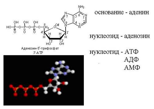
cAMP - "loopback" ATP molecule

Diagram of the nucleotide structure

Note that an activated nucleotide capable of building up a DNA or RNA chain has a "triphosphate tail". It is with this "energy-saturated" tail that it can join the already existing chain of the growing nucleic acid. The phosphate tail sits on carbon 5, so that carbon position is already occupied by phosphates and is meant to be attached. What to attach it to? Only to the carbon at position 3". Once attached, this nucleotide will itself become a target for the next nucleotide to attach. The "receiving side" provides the carbon at position 3", and the "arriving side" clings to it with a phosphate tail located at position 5". In general the chain grows from the 3" side.
Extension of the DNA nucleotide chain
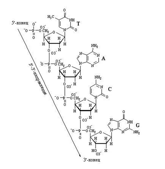
Chain growth due to "longitudinal" bonds between nucleotides can only go in one direction: from 5" ⇒ to 3", because A new nucleotide can only be added to the 3' end of the chain, not to the 5' end.
Pairs of nucleotides connected by "cross" complementary bonds of their nitrogenous bases
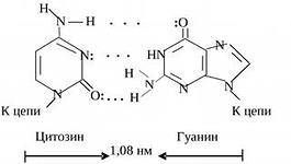
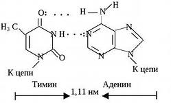
Section of the DNA double helix

Find signs of antiparallelism of two strands of DNA.
Find pairs of nucleotides with double and triple complementary bonds.